Avoid common mistakes on your manuscript.
Hirayama disease is a rare neurological disease, first described by Hirayama in 1959, characterized by insidious unilateral or bilateral muscular atrophy and weakness of the forearms and hands, without sensory or pyramidal signs [1]. The disease affects primarily young men, progresses for a mean of 4–5 years and spontaneously arrests several years after the onset. The majority of the described Hirayama patients are from Asia but cases also from America and Europe have been described. Although its pathophysiological mechanism is still a matter of debate, Kikuchi et al. believe that an imbalanced growth causes disproportion in the lengths of the vertebral column and the spinal canal contents, resulting in a “tight dural sac.”
Cervical MRI in neutral and flexed position is now recommended for the diagnosis since it is easy to perform and reveals various findings on neutral and flexion positioning: localized lower cervical cord atrophy, asymmetric cord flattening, parenchymal changes in the lower cervical cord, abnormal cervical curvature, loss of attachment between the posterior dural sac and subjacent lamina have been described [2]. On flexion MRI, forward migration of the dura mater’s wall is observed with an enlarged posterior epidural space.
We recently demonstrated the ability to identify a metabolic alteration of the spinal cord in patients with motor neuron disease through 18F-Fluorodeoxyglucose Positron Emission Tomography/Computed Tomography (FDG-PET/CT) [3]. In particular, increased metabolic activity has been described at the medulla’s cervical tract level in the presence of Amyotrophic Lateral Sclerosis (ALS). However, no study has ever been published, on spinal metabolism in Hirayama Disease, which can be considered a sort of lower motor neuron disease.
A 16-year-old basketball player presented with a 5-months history of weakness and hypotrophy of the left upper limb’s distal portion.
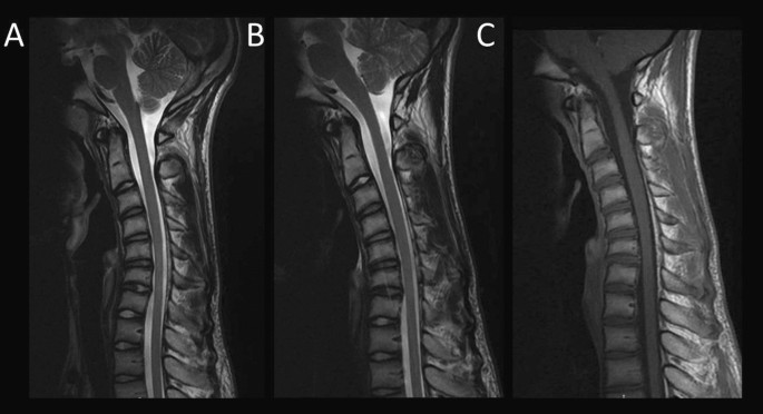
Dynamic cervical MRI revealed, at C5-C6 level, localized lower cervical cord atrophy, asymmetric cord flattening, abnormal cervical curvature and loss of attachment between the posterior dural sac and subjacent lamina in the neutral position and forward migration of the wall of the dura mater in the fully flexed position (Fig. 1). Within 3 months from the dynamic cervical MRI, the patient underwent a total-body FDG-PET/CT scan. Intriguingly, a diffuse, though localized, hypermetabolism was documented in the same cervical tract of the spinal cord (Maximum Standardized Uptake Value, SUVmax 3.5) (Fig. 2). Even though FDG-PET/CT and dynamic MRI images were not superimposed due to different patient positioning, they revealed an appreciable colocalization of the above described pathological findings.
Spinal Cord dynamic MRI alteration: localized lower cervical cord atrophy, asymmetric cord flattening, abnormal cervical curvature and loss of attachment between the posterior dural sac and subjacent lamina in the neutral position (a) and forward migration of the wall of the dura mater with hyperintense enlarged posterior epidural space showing curvilinear flow voids (b) and uniform enhancement after administration of contrast (c) in a fully flexed position
Our studies in ALS patients identified hypermetabolism on the spinal cord, which was extended to the involvement of skeletal muscle, facing an opposite pattern in the brain cortex that showed a generalized reduction in tracer uptake [3]. Of note, in ALS, spinal cord hypermetabolism was able to track disease severity being inversely associated with enrolled patients’ overall survival [3]. Although the exact mechanisms underlying the similar observation in Hirayama disease are probably divergent from ALS, and cannot be exactly identified based on the present data, this finding suggests the possible existence of inflammatory processes involving the cervical tract of the spinal cord in these patients. Currently available anatomopathological studies have shown conflicting results on this topic, agreeing only on the presence of local microvascular alterations. These findings are, specific to this pathology, and certainly different from the results of the anatomopathological studies carried out on ALS patients. However, it cannot be excluded that, as a consequence of different pathogenetic mechanisms, the hypermetabolism shown on FDG-PET may be due to a final inflammatory process common to the two pathologies. Anyway, the colocalization between MRI and PET signals suggests the potential existence of a “metabolic signature” of Hirayama disease (Fig. 3), whose clinical value, given the benign nature of this pathology, needs to be further investigated in the next future, also to increase diagnostic accuracy and to avoid misdiagnosis that could lead to invasive surgical approaches.
Colocalization between the cervical tract hypermetabolism and MRI alterations. Hypermetabolism of the cervical tract of the spinal cord is represented in the Maximum Intensity Projection (a), as well as in the sagittal section (b), while c displays the forward migration of the wall of the dura mater in the fully flexed position in T2-weighted sequence
Data availability
Raw data are available upon appropriate request.
References
Hirayama K (2000) Juvenile Muscular atrophy of distal upper extremity (Hirayama Disease). Intern Med 39(4):283–290
Chen CJ, Hsu HL, Tseng YC, Lyu RK, Chen CM, Huang YC et al (2004) Hirayama flexion myelopathy: neutral position mr imaging findings-importance of loss of attachment. Radiology 231:39–44
Marini C, Morbelli S, Cistaro A, Campi C, Caponnetto C, Bauckneht M, Bellini A, Buschiazzo A, Calamia I, Beltrametti MC, Margotti S, Fania P, Poggi I, Cabona C, Capitanio S, Piva R, Calvo A, Moglia C, Canosa A, Massone A, Nobili F, Mancardi G, Chiò A, Piana M, Sambuceti G (2018) Interplay between spinal cord and cerebral cortex metabolism in amyotrophic lateral sclerosis. Brain 141(8):2272–2279. https://doi.org/10.1093/brain/awy152
Funding
The authors received no financial support for the research.
Author information
Authors and Affiliations
Contributions
BM: Manuscript writing and editing, literature search and review. BL: Data collection, manuscript editing. BA: Data collection, manuscript editing. CC: Data collection, manuscript writing and editing, literature search and review. DS: Data collection, manuscript editing.
Corresponding author
Ethics declarations
Conflicts of interest
None of the authors has any conflict of interest to disclose.
Ethical approval
IRB approved the publication.
Informed consent for publication
Obtained from the patient.
Additional information
Publisher's Note
Springer Nature remains neutral with regard to jurisdictional claims in published maps and institutional affiliations.
Rights and permissions
About this article
Cite this article
Cabona, C., Benedetti, L., Delucchi, S. et al. Cervical alterations in Hirayama disease: an MRI and FDG-PET combined approach. Clin Transl Imaging 9, 117–119 (2021). https://doi.org/10.1007/s40336-021-00409-0
Received:
Accepted:
Published:
Issue Date:
DOI: https://doi.org/10.1007/s40336-021-00409-0


