
Abstract
The aim of the English NHS Diabetic Eye Screening Programme is to reduce the risk of sight loss amongst people with diabetes by the prompt identification and effective treatment if necessary of sight-threatening diabetic retinopathy, at the appropriate stage during the disease process. In order to achieve the delivery of evidence-based, population-based screening programmes, it was recognised that certain key components were required. It is necessary to identify the eligible population in order to deliver the programme to the maximum number of people with diabetes. The programme is delivered and supported by suitably trained, competent, and qualified, clinical and non-clinical staff who participate in recognised ongoing Continuous Professional Development and Quality Assurance schemes. There is an appropriate referral route for those with screen-positive disease for ophthalmology treatment and for assessment of the retinal status in those with poor-quality images. Appropriate assessment of control of their diabetes is also important in those who are screen positive. Audit and internal and external quality assurance schemes are embedded in the service. In England, two-field mydriatic digital photographic screening is offered annually to all people with diabetes aged 12 years and over. The programme commenced in 2003 and reached population coverage across the whole of England by 2008. Increasing uptake has been achieved and the current annual uptake of the programme in 2015–16 is 82.8% when 2.59 million people with diabetes were offered screening and 2.14 million were screened. The benefit of the programme is that, in England, diabetic retinopathy/maculopathy is no longer the leading cause of certifiable blindness in the working age group.
Background
A reduction in diabetes-related blindness by at least one-third was declared a primary objective for Europe in 1989 in the St. Vincent Declaration [1]. Countrywide population-based diabetic retinopathy screening programmes have developed in Iceland (17,200 with diabetes [2] in 2015), Scotland (271,300 people [3] with diabetes), Wales (183,300 people [3] with diabetes), Northern Ireland (84,800 [3] people with diabetes) and England (2.91 million people [3] with diabetes). Regional and local screening programmes have developed in other parts of Europe [4] and around the world. The cost of the English Screening Programme is believed to be approximately 85.6 million US dollars or 40 US dollars per person screened.
The Wilson and Junger criteria for a screening programme, which are the 1968 principles [5] applied by the World Health Organisation, formed the basis of the UK National Screening Committee criteria for appraising the viability, effectiveness and appropriateness of a screening programme when the English NHS Diabetic Eye Screening Programme commenced in 2003. I previously described how we applied these principles to sight-threatening diabetic retinopathy to provide an evidence base [6, 7] for the development of the programme.
It is important to realise the following principles of screening:
-
1.
Screening is a public health programme, not a diagnostic test.
-
2.
Large numbers of apparently healthy individuals are invited for screening, and, if their screening test is positive, offer further diagnostic investigation.
-
3.
Some people may be harmed by the process, or falsely reassured.
-
4.
There is an ethical and moral responsibility to ensure that the programmes are of high quality.
-
5.
Quality Assurance of Screening programmes is therefore essential to ensure that the programme achieves the highest possible standards and minimises harm.
These principles are fundamentally different to most branches of medicine where tests are considered to be diagnostic although, even in the circumstances of diagnostic tests, there will be some false positives and some false negatives.
The sensitivity of a screening test is the percentage of the condition that is correctly detected. If a screening test has a sensitivity of 90%, this means that 1 in 10 is missed. The specificity of a screening test is the percentage of people that one refers unnecessarily. If a screening test is 90% specific, this means 1 in 10 is referred unnecessarily. In 1995, a consensus view was put forward by clinicians at a meeting of the British Diabetic Association in Exeter that a screening test for diabetic retinopathy should have a minimum specificity of 80% and a specificity of 95%. Most studies on screening tests for diabetic retinopathy have achieved over 85% against a recognised reference standard and the specificity target of 95% has been achieved if the numbers with ungradable images are not calculated as test positive [8] but has proved more challenging to achieve when they have been calculated as test positive [9, 10].
It is important that any information that is sent to people who are offered screening tests explains to them that it will not detect all people with the disease and that a small number of people will be referred unnecessarily. It is also important to explain in the literature that a screening test for sight-threatening diabetic retinopathy will not pick up all other eye conditions.
Stages in the development of the English NHS diabetic eye screening programme
When developing the NHS Diabetic Eye Screening Programme in England, we needed to consider 11 different stages that are listed in Table 1.
It was critical for diabetologists, ophthalmologists, public health doctors and optometrists to speak with one voice; otherwise, we would never have established the programme. Assessment and treatment facilities are available in England as part of our National Health Service, but this question becomes much more relevant in developing countries where treatment facilities may not be so readily available. There is no point in screening for sight-threatening diabetic retinopathy if treatment facilities are either not available or inadequate.
In England, everyone has a Primary Care Physician (GP) and so we have to obtain details from Primary Care on those diagnosed with diabetes and everyone has an NHS identifier number. A letter is sent out to everyone with diabetes aged 12 years and over to invite them for a diabetic eye screening appointment once a year. National leaflets have been produced to explain about diabetic retinopathy and about the screening test and what happens if screen-positive diabetic retinopathy is found. We have included information in the leaflet that the screening is not a diagnostic test and hence will only detect at best 90% of sight-threatening diabetic retinopathy and not other eye conditions. There has also been active engagement with patient organisations. There are appropriate exclusion criteria for those who do not need to be invited for screening, e.g. those already under ophthalmology and terminally ill.
Software has been developed to provide a single collated list of people with diabetes and for call recall, screening, grading and audit. In the database, the images are attached to patient details and confidentiality of patient data is a priority. The database is regularly backed up and an IT infrastructure has been established for capture and transmission of images to and from the cameras.
In England, we use non-mydriatic cameras and undertake mydriatic photography on all people with diabetes aged 12 years and older. The two 45° fields captured by the English Screening Programme are shown in Fig. 1 together with the one 45° field used by Scotland and the seven 30° stereo fields that are used as a reference standard against which screening tests are judged. The English Screening Programme sets a minimum camera specification and tests all prospective cameras that meet this minimum specification on patients who are known to have specific features of diabetic retinopathy. The specification document is a fairly lengthy document which includes the following statements: The unit must be capable of providing a minimum field of view of 45° horizontally and 40° vertically at the specified resolution (at least 30 pixels/degree). The unit must be capable of accommodating refractive errors of ±15 D as detailed in EN ISO 10940. The internal fixation aid should be capable of positioning the eye to capture the fields of regard specified below. The ‘field of regard’ of the fundus camera must make it relatively straightforward for an appropriately trained and competent retinal screener to capture images centred on (1) the foveal area and (2) the optic disc. In addition, the ‘field of regard’ of the fundus camera must be able to capture images as defined by the area covered by fields 3–7 of the seven-field protocol used in the Early Treatment Diabetic Retinopathy Study [11]. The list of cameras that are currently approved to be used in the English Screening Programme is published by Public Health England on their webpage [12].

Photographic fields
There has been a progressive increase in the size of uncompressed images from the modern camera backs which are now over 20 MB—the English NHS Diabetic Eye Screening Programme recommends capture of images that are compressed to a size of 1–2 MB. This level of compression has not been shown to lose any clinically significant information [13,14,15].
When considering whether to routinely dilate the pupil of people with diabetes attending for screening the study that was influential in the decision-making process was a population-based screening study [16] of 1549 people with diabetes who had received non-mydriatic one-field digital photography followed by mydriatic two-field digital photography and a reference standard examination by an experienced ophthalmologist whose examination was tested separately against seven-field stereo-photography [17]. The sensitivity for one-field non-mydriatic photography was 86.0% (95% CI, 80.9–91.1%), the specificity was 76.7% (95% CI, 74.5–78.9%) and the poor-quality image rate was 19.7% (95% CI, 18.4–21.0%). The sensitivity for two-field mydriatic photography was 87.8% (95% CI, 83.0–92.6%), the specificity was 86.1% (95% CI, 84.2–87.8%) and a poor-quality image rate was 3.7% (95% CI, 3.1–4.3%). This study led to the approach used in England of two-field mydriatic photography and the approach in Scotland of staged mydriasis with one-field non-mydriatic photography and with dilation only if poor-quality images were obtained. The correlation [18] with age led Northern Ireland to only routinely dilate those aged 50 years and over.
It has been demonstrated that there is a strong correlation [18] between age and poor-quality image rates in diabetic retinopathy screening, for both non-mydriatic and mydriatic photography. Hence, publications [19,20,21,22] with small numbers in a young age range are not relevant to population-based screening programmes where many of the individuals to be screened are over 60 years.
In any population-based screening programme, it is necessary to balance acceptability to the individuals being screened and cost-effectiveness of the screening method with detection rates of sight-threatening diabetic retinopathy. Population-based screening programmes that use non-mydriatic photography like the Scottish Screening Programme [23] usually capture one field centred on the fovea and those that use mydriatic photography like the English Screening Programme usually capture a second field that is centred on the disc, which also give a second view of the macular area. In 1989, Moss [11] demonstrated that for eight retinopathy levels, the rate of agreement with seven stereoscopic fields ranges from 80% for two 30° stereo fields to 91% for four 30° stereo fields. In 2003, Scanlon [17] reported that two-field mydriatic digital photography gave a sensitivity of 80.2% (75.2–85.2) and specificity of 96.2% (93.2–99.2) in comparison with seven-field stereo-photography. In the latter study, 15.3% of seven-field sets were ungradable compared with 1.5% of the two-field digital photographs.
Clear protocols need to be in place for management of people with poor-quality images. In the English Screening Programme, all people with poor-quality images are referred for examination by slit lamp biomicroscopy.
The English NHS Diabetic Eye Screening Programme routinely measures Visual Acuity at screening, but it is recognised that it is not sufficiently sensitive on its own to be a screening tool [24, 25]. Hence, it needs to be used in conjunction with other features that are detected at grading.
The diabetic retinopathy grading classification that has the best evidence base is the Early Treatment Diabetic Retinopathy Study (ETDRS) final Retinopathy Severity Scale [26] because it provided the first detailed classification system for retinopathy severity based on a natural history study of untreated eyes. However, this relies on detailed grading of stereo-photographs of seven fields of each eye. This scale did not grade lesions in the macular area. The ETDRS study did define ‘clinically significant macular oedema’ which was a level at which laser treatment was advised, but this was based on stereo-photography and the study did not recommend a referral level for closer observation before laser treatment was recommended.
Table 2 shows the International Classification [27] which was developed by the American Academy of Ophthalmology in 2002 and recommends that any level of retinopathy more severe than mild retinopathy (defined as the presence of microaneurysms only) warrants examination by an ophthalmologist. However, this is too early a referral level for use in the English Screening Programme and the referral level for the English Screening Programme is also listed in Table 2.
Table 3 shows the risks of progression to proliferative diabetic retinopathy as recorded in the Early Treatment Diabetic Retinopathy Study [26]. Screening Programmes need to accept a certain level of risk. In the English programme, we needed to decide whether we were prepared to accept a 6.2% risk or an 11.3% risk that a patient who has been screened and given a 1-year appointment develops proliferative DR before their next screen. We opted for the 11.3% risk which is the equivalent to moderate non-proliferative diabetic retinopathy on the ETDRS final Retinopathy Severity Scale [26]. We also had to develop a definition for maculopathy referral based on two-dimensional markers. The ETDRS study did not classify maculopathy, but it did make recommendations on what constituted clinically significant macular oedema requiring laser treatment as shown in Table 4. We opted for three referral criteria, based on two-dimensional photographic markers and measurement of Visual Acuity:
-
1.
Exudate within 1 disc diameter (DD) of the centre of the fovea (Fig. 2).
Fig. 2 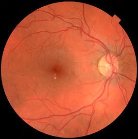
Exudate within 1 disc diameter (DD) of the centre of the fovea
-
2.
Circinate or group of exudates within the macula (Fig. 3).
Fig. 3 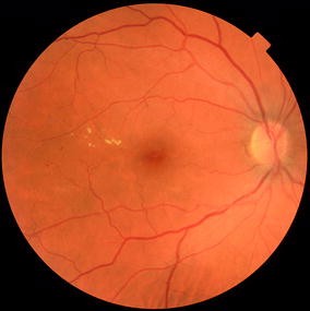
Circinate or group of exudates within the macula
-
3.
Any microaneurysm or haemorrhage within 1DD of the centre of the fovea only if associated with a best VA of ≤ 6/12 (if no stereo) (Fig. 4).
Fig. 4 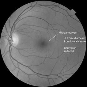
A microaneurysm within 1DD of the centre of the fovea associated with a best VA of ≤ 6/12


A minimum screen resolution is recommended [12] when viewing the images for grading which has progressed as the technology of screens has advanced. The current minimum acceptable standard for screen resolution is a vertical resolution of 1080 (1920 × 1080) with an achievable and recommended standard of a minimum of 1200 (1920 × 1200 or higher). It is recommended that a minimum of 60% of the image should be viewable on the grading screen to avoid too much scrolling to see the full image.
To ensure that whole screening programme is provided by a trained and competent workforce a minimum qualification [28] is required for screeners and graders in the English programme. Evidence of ongoing continuous professional development and taking the monthly External Quality Assurance Test sets [29, 30] is also required—all 1500 graders in the English Screening Programme are required to take a monthly test set of 20 image sets and their grading of these images is compared against a guide grade. An International Version of the qualification [31] and the monthly test and training set [32] for screeners working outside the UK is available.
An important part of any screening programme is the introduction of Quality Assurance. The purpose of introducing Quality Assurance is to reduce the probability of error and risk, ensure that errors are dealt with competently and sensitively, help professionals and organisations improve year on year, and set and keep under review national standards.
The NHS Diabetic Eye Screening Programme has developed three Key Performance Indicators and nine other Quality Standards [33]. These are given in Table 5 with the three Key Performance Indicators being shown in the right-hand column. A programme board which includes local health service representatives and national Quality Assurance team representatives oversees the results of a programmes performance against the standards four times a year, and if a programme is performing poorly, they are expected to improve or the service may be recommissioned to be provided by a different provider. Graders who perform poorly on test sets undergo extra training and have all of their work second graded until their performance improves.
In addition, an External Quality Assurance visit to all regional programmes who undertake Diabetic Eye Screening as part of the NHS Diabetic Eye Screening Programme is undertaken every 3 years. EQA visits are an integral part of Diabetic Eye Screening Quality Assurance. Formal EQA visits to a screening programme provide the forum for a review of the whole multidisciplinary screening pathway and an assessment of the effectiveness of team working within the screening centre and associated referral sites.
Programme results
In the development of the programme, I calculated [7] that the NHS Diabetic Eye Screening Programme had the potential to reduce the prevalence of blindness in England from 4200 people to under 1000 people based on UK certification of blindness. If WHO definitions were used the prevalence, incidence and potential reductions in blindness are much greater. In 2014, Liew [34] reported on the causes of blindness certifications in England and Wales in working age adults (16–64 years) in 2009–2010 and compared these with figures from 1999 to 2000. For the first time in at least five decades, diabetic retinopathy/maculopathy was no longer the leading cause of certifiable blindness amongst working age adults in England and Wales, having been overtaken by inherited retinal disorders. This change was considered to be due to the introduction of nationwide diabetic retinopathy screening programmes in England and Wales and improved glycaemic control. The era in which this reduction in blindness occurred was during the period when laser treatment was being used for maculopathy and before the use of VEGF inhibitors for diabetic macular oedema.
In 2015–2016, the NHS Diabetic Eye Screening Programme in England [35] offered screening to 2,590,082 with diabetes using two-field mydriatic digital photography. There were 3,083,401 known people with diabetes in England, but people who are under an ophthalmologist for diabetic eye disease and certain other categories of people (e.g. terminally ill) are not invited. A total of 2,144,007 with diabetes were screened (Uptake 82.8%). New registrations to programmes in 2015–2016 were 326,587. There were 7593 urgent referrals with proliferative retinopathy and 52,597 referrals with screen-positive maculopathy or pre-proliferative diabetic retinopathy. Rate of retinopathy per 100,000 screened was 2807.
Future developments for the programme
Changes in technology have introduced three-dimensional imaging in the form of Optical Coherence Tomography. These machines are more costly than digital cameras and are not felt to be cost-effective as a first-line screening tool when 65% of the population of people with diabetes have no retinopathy. However, there is a high possibility that they will be introduced as a second-line screening tool for screen-positive maculopathy using two-dimensional markers. It is believed that, of the 52,597 referrals with screen-positive maculopathy, only 20% actually require treatment and a significant proportion of the remaining 80% could be followed up in a technician-led clinic [36] that includes OCT images to exclude any significant diabetic macular oedema. Cost-effectiveness data are needed before this can be introduced.
Extensive work has been done in the area [37,38,39,40] of extended screening intervals for those at low risk. The UK National Screening Committee agreed at their committee on 19 November 2015 and published their recommendation in January 2016 that:
-
(a)
For people with diabetes at low risk of sight loss, the interval between screening tests should change from 1 to 2 years.
-
(b)
The current 1 -year interval should remain unchanged for the remaining people at high risk of sight loss.
The introduction of this extension of screening interval for those with no retinopathy on two consecutive screens, which is the current recommendation in England, is dependent on software development for the programme.
The use of automated analysis is currently being evaluated for use in the English Screening Programme. A recent HTA report [41] has been published on this topic. There are different ways in which automated analysis could be used:
-
(a)
To classify images as no diabetic retinopathy or diabetic retinopathy so that a human grader would only need to look at those with diabetic retinopathy.
-
(b)
To detect referral levels of retinopathy.
-
(c)
To act as a quality assurance tool for retinopathy that is missed.
-
(d)
To determine which images are gradable and which are ungradable.
Scanning laser ophthalmoscopes and wide-field imaging have been widely studied [42,43,44], but this method has not yet been shown to be cost-effective. The earlier devices that provided wide-field imaging compromised [45] on the detection of microaneurysms in the central field.
No hand-held device has ever been shown [46] to have comparable sensitivities and specificities for the detection of sight-threatening diabetic retinopathy to devices where the camera is fixed and the patient’s head is placed on a chin rest and forehead against a fixed band and cannot, therefore, be recommended for population-based screening at the present time.
OCT angiography is new technology [47] that is not currently suitable for population-based screening.
Conclusions
Screening for sight-threatening diabetic retinopathy has been shown to be very effective in England in reducing blindness due to diabetic retinopathy and reducing the number of vitrectomies being performed on advanced disease.
References
Diabetes care and research in Europe (1990) The Saint Vincent declaration. Diabet Med 7:360
Iceland (2015) http://www.idf.org/membership/eur/iceland. Accessed 03 Feb 1017
Facts and Stats (2016) https://www.diabetes.org.uk/Documents/Position%20statements/DiabetesUK_Facts_Stats_Oct16.pdf. Accessed 03 Feb 2017
Screening for diabetic retinopathy in Europe—strategies for overcoming hurdles to progress (2011) http://www.drscreening2005.org.uk/gdansk_2011.html. Accessed 03 Feb 2017
Wilson J, Jungner G (1968) The principles and practice of screening for disease. Public Health Papers 34. Public Health Papers, WHO, Geneva
Scanlon P (2005) An evaluation of the effectiveness and cost-effectiveness of screening for diabetic retinopathy by digital imaging photography & technician ophthalmoscopy & the subsequent change in activity, workload and costs of new diabetic ophthalmology referrals. [M.D.]: London
Scanlon PH (2008) The English national screening programme for sight-threatening diabetic retinopathy. J Med Screen 15:1–4
Pandit RJ, Taylor R (2002) Quality assurance in screening for sight-threatening diabetic retinopathy. Diabet Med 19:285–291
Scanlon P (2005) An evaluation of the effectiveness and cost-effectiveness of screening for diabetic retinopathy by digital imaging photography and technician ophthalmoscopy and the subsequent change in activity, workload and costs of new diabetic ophthalmology referrals. UCL, London
Harding SP, Broadbent DM, Neoh C, Vora J, Williams EMI (1994) The liverpool diabetic eye study—sensitivity and specificity of photography and direct ophthalmoscopy in the detection of sight threatening eye disease. Diabetic Med 79:S45
Moss SE, Meuer SM, Klein R, Hubbard LD, Brothers RJ, Klein BE (1989) Are seven standard photographic fields necessary for classification of diabetic retinopathy? Invest Ophthalmol Vis Sci 30:823–828
Diabetic eye screening: guidance on camera approval (2016). https://www.gov.uk/government/publications/diabetic-eye-screening-approved-cameras-and-settings/diabetic-eye-screening-guidance-on-camera-approval. Accessed 03 Feb 2017
Basu A (2006) Digital image compression should be limited in diabetic retinopathy screening. J Telemed Telecare 12:163–165
Conrath J, Erginay A, Giorgi R et al (2007) Evaluation of the effect of JPEG and JPEG2000 image compression on the detection of diabetic retinopathy. Eye 21:487–493
Li HK, Florez-Arango JF, Hubbard LD, Esquivel A, Danis RP, Krupinski EA (2010) Grading diabetic retinopathy severity from compressed digital retinal images compared with uncompressed images and film. Retina 30:1651–1661
Scanlon PH, Malhotra R, Thomas G et al (2003) The effectiveness of screening for diabetic retinopathy by digital imaging photography and technician ophthalmoscopy. Diabet Med 20:467–474
Scanlon PH, Malhotra R, Greenwood RH et al (2003) Comparison of two reference standards in validating two field mydriatic digital photography as a method of screening for diabetic retinopathy. Br J Ophthalmol 87:1258–1263
Scanlon PH, Foy C, Malhotra R, Aldington SJ (2005) The influence of age, duration of diabetes, cataract, and pupil size on image quality in digital photographic retinal screening. Diabetes Care 28:2448–2453
Massin P, Erginay A, Ben Mehidi A et al (2003) Evaluation of a new non-mydriatic digital camera for detection of diabetic retinopathy. Diabet Med 20:635–641
Cavallerano JD, Aiello LP, Cavallerano AA et al (2005) Non mydriatic digital imaging alternative for annual retinal examination in persons with previously documented no or mild diabetic retinopathy. Am J Ophthalmol 140:667–673
Aptel F, Denis P, Rouberol F, Thivolet C (2008) Screening of diabetic retinopathy: effect of field number and mydriasis on sensitivity and specificity of digital fundus photography. Diabetes Metab 34(3):290–293
Vujosevic S, Benetti E, Massignan F et al (2009) Screening for diabetic retinopathy: 1 and 3 Nonmydriatic 45° digital fundus photographs vs 7 standard early treatment diabetic retinopathy study fields. Am J Ophthalmol 148:111–118
Scottish Diabetic Retinopathy Screening (DRS) Collaborative (2017). http://www.ndrs.scot.nhs.uk/Links/index.htm/. Accessed 04 Jan 2017
Scanlon PH, Foy C, Chen FK (2008) Visual acuity measurement and ocular co-morbidity in diabetic retinopathy screening. Br J Ophthalmol 92:775–778
Corcoran JS, Moore K, Agarawal OP, Edgar DF, Yudkin J (1985) Visual acuity screening for diabetic maculopathy. Practical Diabetes 2:230–232
Early Treatment Diabetic Retinopathy Study Research Group (1991) Fundus photographic risk factors for progression of diabetic retinopathy. ETDRS report number 12. Ophthalmology 98:823–833
Wilkinson CP, Ferris FL 3rd, Klein RE et al (2003) Proposed international clinical diabetic retinopathy and diabetic macular edema disease severity scales. Ophthalmology 110:1677–1682
Continuous professional development for screening—the new qualification (2017). https://cpdscreening.phe.org.uk/healthscreenerqualification. Accessed 04 Jan 2017
Updates to test and training system benefit diabetic eye screening providers (2017). https://phescreening.blog.gov.uk/2016/08/23/updates-to-test-and-training-system-benefit-diabetic-eye-screening-providers/. Accessed 04 Jan 2017
NHS public health functions agreement (2016–17) Service specification no. 22. NHS Diabetic Eye Screening Programme. https://www.england.nhs.uk/commissioning/wp-content/uploads/sites/12/2016/02/serv-spec-22.pdf. Accessed 04 Jan 2017
Certificate of higher education in diabetic retinopathy screening (2017). http://drscreening.org/pages/default.asp?id=2&sID=3. Accessed 04 Jan 2017
International test and training (2017). http://drscreening.org/pages/default.asp?id=27&sID=40. Accessed 04 Jan 2017
Pathway standards for NHS diabetic eye screening programme (2016) https://www.gov.uk/government/uploads/system/uploads/attachment_data/file/543686/Diabetic_eye_screening_pathway_standards.pdf. Accessed 04 Jan 2017
Liew G, Michaelides M, Bunce C (2014) A comparison of the causes of blindness certifications in England and Wales in working age adults (16–64 years), 1999–2000 with 2009–2010. BMJ Open 4:e004015
NHS screening programmes in England. 1 April 2015 to 31 March 2016 (2016). https://www.gov.uk/government/uploads/system/uploads/attachment_data/file/574713/Screening_in_England_2015_to_2016.pdf. Accessed 04 Jan 2017
Mackenzie S, Schmermer C, Charnley A et al (2011) SDOCT imaging to identify macular pathology in patients diagnosed with diabetic maculopathy by a Digital photographic retinal screening programme. PLoS ONE 6:e14811
Stratton IM, Aldington SJ, Taylor DJ, Adler AI, Scanlon PH (2013) A simple risk stratification for time to development of sight-threatening diabetic retinopathy. Diabetes Care 36:580–585
Scanlon PH, Aldington SJ, Leal J et al (2015) Development of a cost-effectiveness model for optimisation of the screening interval in diabetic retinopathy screening. Health Technol Assess 19:1–116
Aspelund T, Thornorisdottir O, Olafsdottir E et al (2011) Individual risk assessment and information technology to optimise screening frequency for diabetic retinopathy. Diabetologia 54:2525–2532
Lund SH, Aspelund T, Kirby P et al (2016) Individualised risk assessment for diabetic retinopathy and optimisation of screening intervals: a scientific approach to reducing healthcare costs. Br J Ophthalmol 100:683–687
(2016) An observational study to assess if automated diabetic retinopathy image assessment software can replace one or more steps of manual imaging grading and to determine their cost-effectiveness. https://www.journalslibrary.nihr.ac.uk/hta/hta20920/#/full-report
Silva PS, Cavallerano JD, Sun JK, Soliman AZ, Aiello LM, Aiello LP (2013) Peripheral lesions identified by mydriatic ultrawide field imaging: distribution and potential impact on diabetic retinopathy severity. Am Acad Ophthalmol 120:2587–2595
Silva PS, Cavallerano JD, Tolls D et al (2014) Potential efficiency benefits of nonmydriatic ultrawide field retinal imaging in an ocular telehealth diabetic retinopathy program. Diabetes Care 37:50–55
Liegl R, Liegl K, Ceklic L et al (2014) Nonmydriatic ultra-wide-field scanning laser ophthalmoscopy (Optomap) versus two-field fundus photography in diabetic retinopathy. Ophthalmologica 231:31–36
Wilson PJ, Ellis JD, MacEwen CJ, Ellingford A, Talbot J, Leese GP (2010) Screening for diabetic retinopathy: a comparative trial of photography and scanning laser ophthalmoscopy. Ophthalmologica 224:251–257
Yogesan K, Constable IJ, Barry CJ, Eikelboom RH, McAllister IL, Tay-Kearney ML (2000) Telemedicine screening of diabetic retinopathy using a hand-held fundus camera. Telemed J 6:219–223
Al-Sheikh M, Akil H, Pfau M, Sadda SR (2016) Swept-source OCT angiography imaging of the foveal avascular zone and macular capillary network density in diabetic retinopathy. Invest Ophthalmol Vis Sci 57:3907–3913
Harding S, Greenwood R, Aldington S et al (2003) Grading and disease management in national screening for diabetic retinopathy in England and Wales. Diabet Med 20:965–971
Treatment Diabetic Retinopathy Study Research Group (1987) Treatment techniques and clinical guidelines for photocoagulation of diabetic macular edema. Early treatment diabetic retinopathy study report number 2. Early Ophthalmol 94:761–774
Author information
Authors and Affiliations
Corresponding author
Ethics declarations
Conflict of interest
None.
Ethical standard
The author has complied with the journals ethical standards.
Statement of human and animal rights
This article does not contain any studies with human or animal subjects performed by the any of the authors.
Informed consent
All patients screened in the English Diabetic Eye Screening Programme provide informed consent to the procedure.
Additional information
Managed by Massimo Porta.
Rights and permissions
Open Access This article is distributed under the terms of the Creative Commons Attribution 4.0 International License (http://creativecommons.org/licenses/by/4.0/), which permits unrestricted use, distribution, and reproduction in any medium, provided you give appropriate credit to the original author(s) and the source, provide a link to the Creative Commons license, and indicate if changes were made.
About this article

Cite this article
Scanlon, P.H. The English National Screening Programme for diabetic retinopathy 2003–2016. Acta Diabetol 54, 515–525 (2017). https://doi.org/10.1007/s00592-017-0974-1
Received:
Accepted:
Published:
Issue Date:
DOI: https://doi.org/10.1007/s00592-017-0974-1