
Abstract
Intercellular communication plays a key role in information processing in the nervous system, to immune response, to cellular growth and differentiation, and to most processes fundamental to life for multicellular organisms. As a sampling technique microdialysis enables in vivo studies of brain and other tissues and has advanced our understanding of intercellular signal processing. The first part of this chapter is an overview of microdialysis in the context of a how-to-guide with reference to sampling extrasynaptic glutamate and GABA using conventional methodology. The limitations and challenges associated with sampling the synaptic pool of fast neurotransmitters are then addressed. The last part of this chapter presents ideas of advancing the microdialysis technique that may bring the microdialysis membrane closer to the synapse.
Access this chapter
Tax calculation will be finalised at checkout
Purchases are for personal use only
References
Herrera-Marschitz M, Arbuthnott G, Ungerstedt U (2010) The rotational model and microdialysis: significance for dopamine signalling, clinical studies, and beyond. Prog Neurobiol 90:176–189
Dupre KB, Ostock CY, Eskow Jaunarajs KL et al (2011) Local modulation of striatal glutamate efflux by serotonin 1A receptor stimulation in dyskinetic, hemiparkinsonian rats. Exp Neurol 229(2):288–299
Lönnroth P, Jansson PA, Smith U (1987) A microdialysis method allowing characterization of intercellular water space in humans. Am J Physiol 253:E228–E231
Willuhn I, Wanat MJ, Clark JJ et al (2010) Dopamine signaling in the nucleus accumbens of animals self-administering drugs of abuse. Curr Top Behav Neurosci 3:29–71
Zetterstrom T, Sharp T, Marsden CA et al (1983) In vivo measurement of dopamine and its metabolites by intracerebral dialysis: changes after d-amphetamine. J Neurochem 41:1769–1773
Bito L, Davson H, Levin E et al (1966) The concentrations of free amino acids and other electrolytes in cerebrospinal fluid, in vivo dialysate of brain and blood plasma of the dog. J Neurosci 13:1057–1067
Bourne J (2003) Intracerebral microdialysis: 30 Years as a tool for the neuroscientist. Clin Exp Pharmacol Physiol 30:16–24
Benjamin RK, Hochberg FH, Fox E et al (2004) Review of microdialysis in brain tumors, from concept to application: first annual carolyn Frye-Halloran symposium. Neuro Oncol 6(1):65–74
Johansen MJ, Newman RA, Madden T (1997) The use of microdialysis in pharmacokinetics and pharmacodynamics. Pharmacotherapy 17:464–481
Wang PC, DeVoe DL, Lee CS (2001) Integration of polymeric membranes with microfluidic networks for bioanalytical applications. Electrophoresis 22:3857–3867
Páez X, Hemández L (2001) Biomedical applications of capillary electrophoresis with laser-induced fluorescence detection. Biopharm Drug Dispos 22:273–289
Watson CJ, Venton BJ, Kennedy RT (2006) In vivo measurements of neurotransmitters by microdialysis sampling. Anal Chem 78(5):1391–1399
Garrison KE, Pasas SA, Cooper JD et al (2002) A review of membrane sampling from biological tissues with applications in pharmacokinetics, metabolism and pharmacodynamics. Eur J Pharm Sci 17:1–12
Dash MB, Douglas CL, Vyazovskiy VV et al (2009) Long-term homeostasis of extracellular glutamate in the rat cerebral cortex across sleep and waking states. J Neurosci 29:620–629
Drew KL, Pehek EA, Rasley BT et al (2004) Sampling glutamate and GABA with microdialysis: suggestions on how o get the dialysis membrane closer to the synapse. J Neurosci Methods 140:127–131
Bordji K, Becerril-Ortega J, Nicole O et al (2010) Activation of extrasynaptic, but not synaptic, NMDA receptors modifies amyloid precursor protein expression pattern and increases amyloid-ss production. J Neurosci 30:15927–15942
Okubo Y, Iino M (2011) Visualization of glutamate as a volume transmitter. J Physiol 589:481–488
Zhou F, Braddock JF, Hu Y et al (2002) Microbial origin of glutamate, hibernation and tissue trauma: an in vivo microdialysis study. J Neurosci Methods 119:121–128
Sam PM, Justice JB Jr (1996) Effect of general microdialysis-induced depletion on extracellular dopamine. Anal Chem 68:724–728
Langemann H, Alessandri B, Mendelowitsch A et al (2001) Extracellular levels of glucose and lactate measured by quantitative microdialysis in the human brain. Neurol Res 23:531–536
Kennedy RT, Thompson JE, Vickroy TW (2002) In vivo monitoring of amino acids by direct sampling of brain extracellular fluid at ultralow flow rates and capillary electrophoresis. J Neurosci Methods 114:39–49
Osborne PG, Hu Y, Covey DN et al (1999) Determination of striatal extracellular gamma-aminobutyric acid in non-hibernating and hibernating arctic ground squirrels using quantitative microdialysis. Brain Res 839:1–6
Justice JB Jr (1993) Quantitative microdialysis of neurotransmitters. J Neurosci Methods 48:263–276
Parsons LH, Justice JB Jr (1994) Quantitative approaches to in vivo brain microdialysis. Crit Rev Neurobiol 8(3):189–220
Pehek EA, Nocjar C, Roth BL et al (2006) Evidence for the preferential involvement of 5-HT2A serotonin receptors in stress- and drug-induced dopamine release in the rat medial prefrontal cortex. Neuropsychopharmacology 31:265–277
Liu Y, Zhang J, Xu X et al (2010) Capillary ultrahigh performance liquid chromatography with elevated temperature for sub-one minute separations of basal serotonin in submicroliter brain microdialysate samples. Anal Chem 82:9611–9616
Kaul S, Faiman MD, Lunte CE (2011) Determination of GABA, glutamate and carbamathione in brain microdialysis samples by capillary electrophoresis with fluorescence detection. Electrophoresis 32:284–291
Kirschner D, Jaramillo M, Green TK (2007) Enantio separation and stacking of cyanobenz(f)isoindole-amino acids by reverse polarity capillary electrophoresis and sulfated β-cyclodextrin. Anal Chem 79:736–743
Kirschner DL, Green T (2009) Separation and sensitive detection of D-amino acids in biological matrices. J Sep Sci 32:2305–2318
Nandi P, Lunte SM (2009) Recent trends in microdialysis sampling integrated with conventional and microanalytical systems for monitoring biological events: a review. Anal Chim Acta 651:1–14
Carrozzo MM, Cannazza G, Pinetti D et al (2010) Quantitative analysis of acetylcholine in rat brain microdialysates by liquid chromatography coupled with electrospray ionization tandem mass spectrometry. J Neurosci Methods 194:87–93
Dahlin AP, Wetterhall M, Caldwell KD et al (2010) Methodological aspects on microdialysis protein sampling and quantification in biological fluids: an in vitro study on human ventricular CSF. Anal Chem 82:4376–4385
Glick SD, Dong N, Keller RW Jr et al (1994) Estimating extracellular concentrations of dopamine and 3,4-dihydroxyphenylacetic acid in nucleus accumbens and striatum using microdialysis: relationships between in vitro and in vivo recoveries. J Neurochem 62:2017–2021
Wang L, Li Y, Han H et al (2003) Perfusate oxygen and carbon dioxide concentration influence basal microdialysate levels of striatal glucose and lactate in conscious rats. Neurosci Lett 344:91–94
Osborne PG, O’Connor WT, Ungerstedt U (1991) Effect of varying the ionic concentration of a microdialysis perfusate on basal striatal dopamine levels in awake rats. J Neurochem 56:452–456
Oz G, Berkich DA, Henry PG et al (2004) Neuroglial metabolism in the awake rat brain: CO2 fixation increases with brain activity. J Neurosci 24:11273–11279
Duo J, Stenken JA (2011) In vitro and in vivo affinity microdialysis sampling of cytokines using heparin-immobilized microspheres. Anal Bioanal Chem 399:783–793
Benveniste H, Drejer J, Schousboe A et al (1987) Regional cerebral glucose phosphorylation and blood flow after insertion of a microdialysis fiber through the dorsal hippocampus in the rat. J Neurochem 49:729–734
Fumero B, Guadalupe T, Valladares F et al (1994) Fixed versus removable microdialysis probes for in vivo neurochemical analysis: implications for behavioral studies. J Neurochem 63:1407–1415
Osborne PG (1995) Fixed versus removable microdialysis probes for in vivo neurochemical analysis: implications for behavioral studies. J Neurochem 64:1899–1901
Drew KL, O’Connor WT, Kehr J et al (1989) Characterization of gamma-aminobutyric acid and dopamine overflow following acute implantation of a microdialysis probe. Life Sci 45:1307–1317
Timmerman W, Westerink BH (1997) Brain microdialysis of GABA and glutamate: what does it signify? Synapse 27:242–261
Baker DA, Shen H, Kalivas PW (2002) Cystine/glutamate exchange serves as the source for extracellular glutamate: modifications by repeated cocaine administration. Amino Acids 23:161–162
Richerson GB (2004) Looking for GABA in all the wrong places: the relevance of extrasynaptic GABA(A) receptors to epilepsy. Epilepsy Curr 4:239–242
Wu Y, Wang W, Diez-Sampedro A et al (2007) Nonvesicular inhibitory neurotransmission via reversal of the GABA transporter GAT-1. Neuron 56:851–865
Augustin H, Grosjean Y, Chen K et al (2007) Nonvesicular release of glutamate by glial xCT transporters suppresses glutamate receptor clustering in vivo. J Neurosci 27:111–123
Amen SL, Piacentine LB, Ahmad ME et al (2011) Repeated N-acetyl cysteine reduces cocaine seeking in rodents and craving in cocaine-dependent humans. Neuropsychopharmacology 36:871–878
Stanika RI, Pivovarova NB, Brantner CA et al (2009) Coupling diverse routes of calcium entry to mitochondrial dysfunction and glutamate excitotoxicity. Proc Natl Acad Sci U S A 106:9854–9859
Sarrafzadeh A, Haux D, Plotkin M et al (2005) Bedside microdialysis reflects dysfunction of cerebral energy metabolism in patients with aneurysmal subarachnoid hemorrhage as confirmed by 15 O-H2 O-PET and 18 F-FDG-PET. J Neuroradiol 32:348–351
Sarrafzadeh AS, Nagel A, Czabanka M et al (2010) Imaging of hypoxic-ischemic penumbra with (18)F-fluoromisonidazole PET/CT and measurement of related cerebral metabolism in aneurysmal subarachnoid hemorrhage. J Cereb Blood Flow Metab 30:36–45
Herman MA, Jahr CE (2007) Extracellular glutamate concentration in hippocampal slice. J Neurosci 27:9736–9741
Barbour B (2001) An evaluation of synapse independence. J Neurosci 21:7969–7984
Cragg SJ, Rice ME (2004) Dancing past the DAT at a DA synapse. Trends Neurosci 27:270–277
Schonfuss D, Reum T, Olshausen P et al (2001) Modeling constant potential amperometry for investigations of dopaminergic neurotransmission kinetics in vivo. J Neurosci Methods 112:163–172
Nicholson C (2001) Diffusion and related transport mechanisms in brain tissue. Rep Prog Phys 64:815–884
Nicholson C (2005) Factors governing diffusing molecular signals in brain extracellular space. J Neural Transm 112:29–44
Chen KC (2006) Effects of tissue trauma on the characteristics of microdialysis zero-net-flux method sampling neurotransmitters. J Theor Biol 238:863–881
Clapp-Lilly KL, Roberts RC, Duffy LK et al (1999) An ultrastructural analysis of tissue surrounding a microdialysis probe. J Neurosci Methods 90:129–142
Connelly CA (1999) Microdialysis update: optimizing the advantages. J Physiol 514(Pt 2):303
Bungay PM, Newton-Vinson P, Isele W et al (2003) Microdialysis of dopamine interpreted with quantitative model incorporating probe implantation trauma. J Neurochem 86:932
Yang H, Peters JL, Allen C et al (2000) A theoretical description of microdialysis with mass transport coupled chemical events. Anal Chem 72:2042–2049
Chen KC (2005) Preferentially impaired neurotransmitter release sites not their discretiness compromise the validoty of microdialysis zero-net-flux method. J Neurochem 92:29–45
Allen C, Peters JL, Sesack SR et al (2001) Microelectrodes closely approach intact nerve terminals in vivo, while larger devices do not: a study using electrochemistry and electron microscopy. Monitoring molecules in neuroscience.In: Proceedings of the international conference on in vivo methods, 9th. Dublin, Ireland 89–90
Jacobson I, Sandberg M, Hamberger A (1985) Mass transfer in brain dialysis devices–a new method for estimation of extracellular amino acids concentration. J Neurosci Methods 15(3):263–268
Lada MW, Vickroy TW, Kennedy RT (1997) High temporal resolution monitoring of glutamate and aspartate in vivo using microdialysis on-line with capillary electrophoresis with laser-induced fluorescence detection. Anal Chem 69:4560–4565
Lada MW, Vickroy TW, Kennedy RT (1998) Evidence for neuronal origin and metabotropic receptor-mediated regulation of extracellular glutamate and aspartate in rat striatum in vivo following electrical stimulation of the prefrontal cortex. J Neurochem 70:617–625
Wang M, Roman GT, Perry ML et al (2009) Microfluidic chip for high efficiency electrophoretic analysis of segmented flow from a microdialysis probe and in vivo chemical monitoring. Anal Chem 81:9072–9078
Croce G, D’Agaro P (2005) Numerical simulation of roughness effect on microchannel heat transfer and pressure drop in laminar flow. J Phys D: Appl Phys 38:1518–1530
Nguyen N-T, Huang X, Chuan TK (2002) MEMS-micropumps: a review. J Fluids Eng 124:384–392, ASME
Chen C, Drew KL (2008) Droplet-based microdialysis concept, theory, and design consideration. J Chromatogr A 1209:29–38
Bungay PM, Morrison PF, Dedrick RL (1990) Steady state theory for quantitative microdialysis of solutes and water in vivo and in vitro. Life Sci 46:105–119
Maidment NT, Evans CJ (1991) Measurement of intracellular neuropeptides in the brain: microdialysis linked to solid-phase radioimmunoassays with sub-femtomole limits of detection. In: Robinson TE (ed) Microdialysis in the Neurosciences. Elsevier, Amsterdam
Çengel YA, Cimbala JM (2006) Fluid Mechanics fundamentals and applications. McGraw Hill, New York
Taylor RI (1953) Dispersion of soluble matter in solvent flowing slowly through a tube. Proc R Soc Lond A 219:186–203
Yang H, Nguyen NT, Huang X (2006) Micromixer based on Taylor disperson. J Phys 34:136–141, Conference series
Tan J, Li SW, Wang K et al (2009) Gas-liquid flow in T-junction microfluidic devices with a new perpendicular rupturing flow route. Chem Eng J 146:428–433
Teh S-Y, Lin R, Hung LH et al (2008) Droplet microfluidics. Lab Chip 8:198–220
Chen C-S, Breslauer DN, Luna JI et al (2008) Shrinky-Dink microfluidics: 3D polystyrene chips. Lab Chip 8:622–624
Xia Y, Whitesides GM (1998) Soft lithography. Angew Chem Int Ed 37:550–575
Hsieh Y-C, Zahn JD (2007) On-chip microdialysis system with flow-through sensing components. Biosens Bioelectron 22:2422–2428
Polston JE, Rubbinaccio HY, Morra JT et al (2011) Music and methamphetamine: conditioned cue-induced increases in locomotor activity and dopamine release in rats. Pharmacol Biochem Behav 98:54–6182.Shim, J.-U., G. Cristobal, D. Link, T. Thorsen, and S. Fraden (2007). Using microfluidics to decouple nucleation and growth of protein crystals. Cryst Growth Des 7, no. 11 2192–94.
Author information
Authors and Affiliations
Corresponding author
Editor information
Editors and Affiliations
Construction of Low Dead Volume Microdialysis Probes
Construction of Low Dead Volume Microdialysis Probes
-
1.
Prepare capillary: Using a ceramic capillary tubing cutter, cut up to a 50 cm piece of capillary tubing1 (this will be the inlet to the probe, so adjust length as needed). Measure and cut another piece for the outlet. For acute, anesthetized preps this may be 10 cm. For awake animal preparations leave the outlet long enough to reach your fraction collector or collection vial. Note that longer outlets will decrease the flow rate that a probe can handle before breaking or seeping fluid (ultrafiltration). Ensure that cuts are smooth; check smoothness using a microscope. Smoothness means that the tube is not shattered and the cut does not leave a point on the tube.
-
2.
Adjust offset between inlet and outlet that will define the length of dialyzing tip: On a glass slide with double-sided tape on the topside of the slide, lay the long capillary piece on the slide and next to it lay the short capillary piece. Bring the ends of the two pieces flush with one another, and then make the long capillary tube stick out further than the short capillary tube by 1–4 mm or longer depending on the desired length of your dialyzing tip. Put this in an area where it will not be disturbed.
-
3.
Prepare membrane for sliding onto capillaries: Cut 0.5 cm of cellulose membrane2 from the roll at a 45° angle. Put the cut membrane on a glass slide with double-sided tape on the top. Using polyimide resin3, seal the cut membrane end that is not angled. The sealed part should not exceed a thickness of 0.5 mm (Fig. 7a). Leave this to dry for several hours.
Fig. 7. 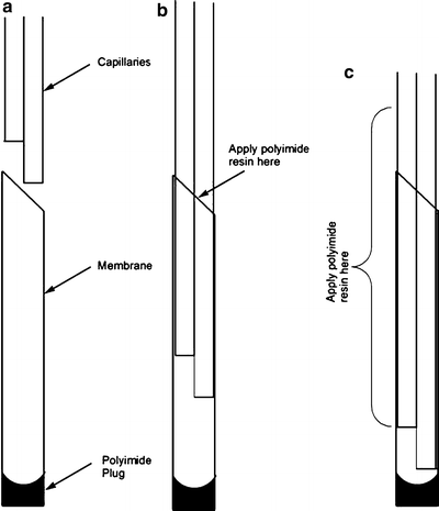
Offset short and long piece of capillary to define the length of dialyzing tip (a). Feed into membrane, sealed on one end with polyimide resin (plug not to exceed 0.5 mm) and trimmed at a 45° angle on the other end. After inserting the capillary tubes into the membrane about 1 mm, apply polyimide resin to the capillaries at the opening of the membrane (b). Coat the membrane and capillary tubes with polyimide resin as shown leaving the dialyzing tip free of resin (c). These membrane-covered capillaries are now referred to as “the probe”.
-
4.
Insert capillary tubes into membrane: Insert offset capillary tubes into a dry, sealed membrane tube. For easier insertion put the short tube on the long side of the angled membrane opening (Fig. 7b).
-
5.
Coat non-dialyzing surface of membrane and define the final length of dialyzing tip: After inserting capillary tubes about 1 mm apply polyimide resin onto the capillaries and to the open end of the membrane (the end cut to 45°). Continue feeding the capillaries into the membrane. Adjust the capillaries so that they remain offset as before, but the long capillary is about 0.1 mm from the polyimide plug at the end of the membrane. Spread polyimide resin on the membrane entrance and the membrane itself. Stop spreading resin just before the opening of the furthest back capillary tube. Remove excess polyimide resin and let dry for several hours (Fig. 7c).
-
6.
Insert the completed probe into a push–pull cannula assembly: The assembly consists of two pieces, the push–pull connector-no internal tube referred to here as the “cap” (C313ICP/NIT4 where NIT indicates “no internal tube”), and the push–pull guide cannula (C316GPIO/uncut4). Attach a 1 cm piece of Tygon tubing (0.020–0.025” id) to the stainless steel tube on the top of the cap that leads to a hole directly in the middle of the cap (Fig. 8a). Thread the probe through this piece of Tygon and stainless steel tube. Next insert the probe through the push–pull guide cannula and then screw the two pieces together for a tight, reproducible fit.
Fig. 8. 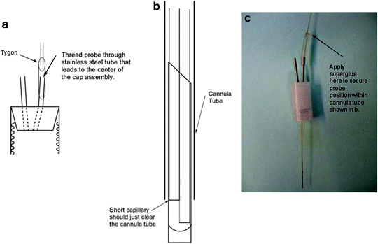
The probe is threaded through a piece of Tygon tubing affixed to the stainless steel tube that leads to the center of the “cap” that is the push–pull connector-no internal tube (Cat No. C313ICP/NIT) (a). The probe that is now connected to the cap is then threaded through the push–pull guide cannula (Cat No. C316GP/O) and the two pieces are screwed together and tightened for a reproducible fit. Adjust the position of the probe within the cannula assembly so that the short capillary just clears the cannula tip and the active portion of the membrane extends beyond the tip (b). Apply a dab of super glue where the probe enters the Tygon tubing (c) and let dry for at least 20 min.
-
7.
Adjust and secure probe within cannula assembly: With the capillary mounted on sticky tack, adjust the push–pull cannula assembly so that active portion of the probe membrane extends just beyond the end of the cannula tip (Fig. 8b). When this is done, apply a little bit of super glue to the entrance of the Tygon to hold the probe in position. Let this dry for 20 min. The fully assembled probe is shown in Fig. 8c and Fig. 9.
Fig. 9. 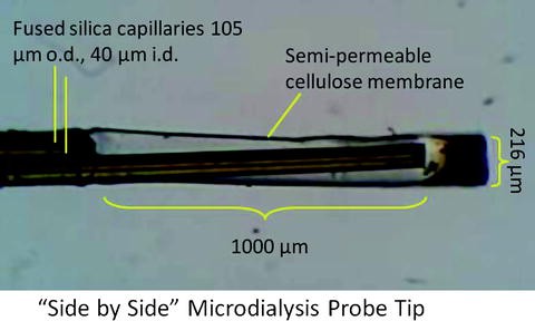
Photograph of dialyzing tip of an assembled probe.
-
8.
Prepare capillary inlet to connect to a syringe filled with perfusion fluid: To fit the capillary tubing onto a luer lock syringe you need to first put the capillary through the luer adapter fitting and then insert the capillary through a red NanoTight®sleeve5. After inserting the capillary through the red NanoTight® sleeve, pull the tubing a little further out and cut off about 3–4 cm of tubing to get rid of capillary tubing that might be obstructed, leaving 1 mm projecting from the red NanoTight® sleeve. Place a ferrule on the sleeve and connect using normal HPLC fittings.
-
9.
Prepare cannula to fit probe holder needed for stereotaxic surgery: If probes will be used in an acute, anesthetized preparation, unscrew the cap and use a dremel tool to file the edges of the outer threads of the internal cannula to make two parallel, flat surfaces. Use a CMA clip designed to hold the flat body of a CMA 11 or 12 guide cannula6 for surgery. If probes will be used for an awake preparation that requires chronic guide cannula implantation, use a nut that threads onto the top of the guide cannula. Flatten the sides of the nut so that it fits securely within the CMA clip. To insure accurate depth of placement take care to adjust the length of capillary and dialyzing membrane to an exact length that is consistent for all probes.
-
10.
Notes on performance: Due to the low internal diameter of the outlet capillary, these probes will sweat at relatively slow flow rates. Probes with short outlets (15 cm) sweat between 1.0 and 8.0 μL/min (mean of 4.7 μL/min, n = 8). Probes with long outlets (100 cm) sweat between 0.3 and 0.8 μL/min (mean of 0.6 μL/min, n = 8). In vitro tests show that when these probes are perfused at 0.1 μL/min the temporal resolution, or time to equilibrate to a new concentration of glu ranging from 0.5 to 2.0 μM, is about 1 min.


1.1 Supply List for Microdialysis Probe Construction
Manufacturer-Specific Items
-
1.
Silica capillary Tubing; 105 μm od, 40 μm id (Polymicro Technologies; http://www.polymicro.com).
Part No. TSP040105.
-
2.
Regenerated cellulose membrane; 13 kDa cut-off (Spectrum Labs; http://www.spectrumlabs.com).
Part No.132294.
-
3.
Polyimide sealing resin; part No.5825 (W.R. Grace and Co.; http://www.discoverysciences.com).
-
4.
Push–pull cannula and cannula guides (Plastics 1; http://www.plastics1.com).
-
Part No.C313ICP/NIT Push–pull connector-internal cannula.
-
Part No. C316GPIO/uncut (maximum length) push–pull guide cannula.
-
-
5.
HPLC fittings and sleeves (Upchurch Scientific; http://www.upchurch.com).
-
Part No. F-237x NanoTight FEP sleeve (0.005”; red).
-
Part No. F-142Nx HPLC ferrule.
-
Part No. F-331Nx HPLC fitting.
-
-
6.
CMA 11 and 12 clip (CMA Microdialysis AB; http://www.microdialysis.se).
General Items (Found in Hardware Store or Scientific Supply Catalog)
-
Small Tygon Tubing (0.020–0.025” id).
-
Super Glue.
-
Epoxy Glue.
-
2-sided tape.
-
Microscope slides.
-
Ceramic capillary tubing cutter.
-
Poster putty.
Rights and permissions
Copyright information
© 2013 Springer Science+Business Media, LLC
About this protocol
Cite this protocol
Chen, Cf., Rasley, B.T., Warlick, B.P.E., Green, T.K., Swearingen, K.E., Drew, K.L. (2013). Microdialysis and Advances for Sampling Synaptic and Extrasynaptic Pools. In: Di Giovanni, G., Di Matteo, V. (eds) Microdialysis Techniques in Neuroscience. Neuromethods, vol 75. Humana Press, Totowa, NJ. https://doi.org/10.1007/978-1-62703-173-8_4
Download citation
DOI: https://doi.org/10.1007/978-1-62703-173-8_4
Published:
Publisher Name: Humana Press, Totowa, NJ
Print ISBN: 978-1-62703-172-1
Online ISBN: 978-1-62703-173-8
eBook Packages: Springer Protocols