Can radiomics improve the prediction of metastatic relapse of myxoid/round cell liposarcomas? Amandine CrombéFrançois Le LoarerMichèle Kind Oncology 17 January 2020 Pages: 2413 - 2424
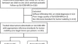
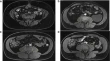
Added value of MRI to endoscopic and endosonographic response assessment after neoadjuvant chemoradiotherapy in oesophageal cancer Sophie E. VollenbrockJolanda M. van DierenAnnemarieke Bartels-Rutten Oncology 21 January 2020 Pages: 2425 - 2434
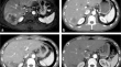
Hepatic hemangioendothelioma: CT, MR, and FDG-PET-CT in 67 patients—a bi-institutional comprehensive cancer center review Dhakshinamoorthy GaneshanPerry J. PickhardtKhaled M. Elsayes Oncology 30 January 2020 Pages: 2435 - 2442
Preoperative 18F-FDG PET/CT tumor markers outperform MRI-based markers for the prediction of lymph node metastases in primary endometrial cancer Kristine E. FasmerAnkush GulatiIngfrid S. Haldorsen Oncology Open access 07 February 2020 Pages: 2443 - 2453
Microwave ablation after downstaging of hepatocellular carcinoma: outcome was similar to tumor within Milan criteria Feng ShiShanshan LianXiaoming Chen Interventional 30 January 2020 Pages: 2454 - 2462
Minimal ablative margin (MAM) assessment with image fusion: an independent predictor for local tumor progression in hepatocellular carcinoma after stereotactic radiofrequency ablation Gregor LaimerPeter SchullianReto Bale Interventional Open access 30 January 2020 Pages: 2463 - 2472
The Focused Ultrasound Myoma Outcome Study (FUMOS); a retrospective cohort study on long-term outcomes of MR-HIFU therapy Inez M. VerpalenJolien P. de BoerManon N. G. Braat Interventional 10 February 2020 Pages: 2473 - 2482
Value of integrated PET-IVIM MR in assessing metastases in hypermetabolic pelvic lymph nodes in cervical cancer: a multi-parameter study Chen XuSiyao DuHongzan Sun Nuclear Medicine 10 February 2020 Pages: 2483 - 2492
Multinational data on cumulative radiation exposure of patients from recurrent radiological procedures: call for action Marco BrambillaJenia VassilevaMadan M. Rehani Computed Tomography 02 December 2019 Pages: 2493 - 2501
Quantitative CT detects progression in COPD patients with severe emphysema in a 3-month interval Philip KonietzkeMark O. WielpützOliver Weinheimer Computed Tomography 21 January 2020 Pages: 2502 - 2512
Pancreatic ductal adenocarcinoma: a radiomics nomogram outperforms clinical model and TNM staging for survival estimation after curative resection Tiansong XieXuanyi WangZhengrong Zhou Computed Tomography 31 January 2020 Pages: 2513 - 2524
The influence of image quality on diagnostic performance of a machine learning–based fractional flow reserve derived from coronary CT angiography Peng Peng XuJian Hua LiLong Jiang Zhang Computed Tomography 31 January 2020 Pages: 2525 - 2534
Quantitative lobar pulmonary perfusion assessment on dual-energy CT pulmonary angiography: applications in pulmonary embolism Ramandeep SinghRyan Zipan NieMannudeep K. Kalra Computed Tomography 31 January 2020 Pages: 2535 - 2542
Carotid near-occlusion is often overlooked when CT angiography is assessed in routine practice Elias JohanssonThomas GuAllan J. Fox Computed Tomography Open access 31 January 2020 Pages: 2543 - 2551
Physical evaluation of an ultra-high-resolution CT scanner Luuk J. OostveenKirsten L. BoedekerIoannis Sechopoulos Computed Tomography Open access 10 February 2020 Pages: 2552 - 2560
Six DWI questions you always wanted to know but were afraid to ask: clinical relevance for breast diffusion MRI Mami IimaSavannah C. PartridgeDenis Le Bihan Magnetic Resonance 21 January 2020 Pages: 2561 - 2570
MRI in patients with implanted active devices: how to combine safety and image quality using a limited transmission field? Laura LundenStephan WolffOlav Jansen Magnetic Resonance 23 January 2020 Pages: 2571 - 2582
MRI of non-specific low back pain and/or lumbar radiculopathy: do we need T1 when using a sagittal T2-weighted Dixon sequence? Fabio ZanchiRaphaël RichardPatrick Omoumi Magnetic Resonance Open access 04 February 2020 Pages: 2583 - 2593
Texture analysis of deep medullary veins on susceptibility-weighted imaging in infants: evaluating developmental and ischemic changes Hyun Gi KimJin Wook ChoiHye Sun Lee Magnetic Resonance 05 February 2020 Pages: 2594 - 2603
Society of Abdominal Radiology (SAR) and European Society of Urogenital Radiology (ESUR) joint consensus statement for MR imaging of placenta accreta spectrum disorders Priyanka JhaLiina PōderGabriele Masselli Magnetic Resonance 10 February 2020 Pages: 2604 - 2615
The prognostic value of late gadolinium enhancement in myocarditis and clinically suspected myocarditis: systematic review and meta-analysis Fuyao YangJie WangYucheng Chen Magnetic Resonance 10 February 2020 Pages: 2616 - 2626
CT and MR imaging prior to transcatheter aortic valve implantation: standardisation of scanning protocols, measurements and reporting—a consensus document by the European Society of Cardiovascular Radiology (ESCR) Marco FranconeRicardo P. J. BuddeRodrigo Salgado Cardiac Open access 05 September 2019 Pages: 2627 - 2650
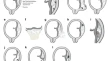
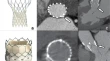
Surgically implanted aortic valve bioprostheses deform after implantation: insights from computed tomography Marguerite E. FaureDominika SucháRicardo P. J. Budde Cardiac 30 January 2020 Pages: 2651 - 2657
Visual scoring of aortic valve calcifications on low-dose CT in lung cancer screening Yeqing ZhuYong WangClaudia I. Henschke Cardiac 10 February 2020 Pages: 2658 - 2668
Stratification of long-term outcome in stable idiopathic pulmonary fibrosis by combining longitudinal computed tomography and forced vital capacity Nicola SverzellatiMario SilvaBrian J. Bartholmai Chest 31 January 2020 Pages: 2669 - 2679
Preoperative CT-based radiomics combined with intraoperative frozen section is predictive of invasive adenocarcinoma in pulmonary nodules: a multicenter study Guangyao WuHenry C. WoodruffPhilippe Lambin Chest Open access 31 January 2020 Pages: 2680 - 2691
Microwave ablation plus chemotherapy versus chemotherapy in advanced non-small cell lung cancer: a multicenter, randomized, controlled, phase III clinical trial Zhigang WeiXia YangHui Tian Chest 04 February 2020 Pages: 2692 - 2702
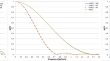
Airway tapering: an objective image biomarker for bronchiectasis Wieying KuoAdria Perez-Roviraon behalf of the Normal Chest CT study group Chest Open access 05 February 2020 Pages: 2703 - 2711
Acquisition time, radiation dose, subjective and objective image quality of dual-source CT scanners in acute pulmonary embolism: a comparative study Waleed AbdellatifEric EsslingerSavvas Nicolaou Chest 05 February 2020 Pages: 2712 - 2721
Chest CT imaging features for prediction of treatment response in cryptogenic and connective tissue disease–related organizing pneumonia Young Hoon ChoEun Jin ChaeSe Jin Jang Chest 10 February 2020 Pages: 2722 - 2730
Cone-beam breast CT features associated with HER2/neu overexpression in patients with primary breast cancer Yueqiang ZhuYuwei ZhangZhaoxiang Ye Breast 03 January 2020 Pages: 2731 - 2739
Comparison of propagation-based CT using synchrotron radiation and conventional cone-beam CT for breast imaging Seyedamir Tavakoli TabaPatrycja BaranPatrick C. Brennan Breast 23 January 2020 Pages: 2740 - 2750
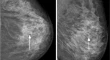
Can breast MRI accurately exclude malignancy in mammographic architectural distortion? Yoav AmitaiAnabel ScaraneloVivianne Freitas Breast 30 January 2020 Pages: 2751 - 2760
Combination of an ultrafast TWIST-VIBE Dixon sequence protocol and diffusion-weighted imaging into an accurate easily applicable classification tool for masses in breast MRI Sandra C. PeterEvelyn WenkelStephan Ellmann Breast 30 January 2020 Pages: 2761 - 2772
BI-RADS category 3, 4, and 5 lesions identified at preoperative breast MRI in patients with breast cancer: implications for management Si Eun LeeJi Hye LeeVivian Youngjean Park Breast 31 January 2020 Pages: 2773 - 2781
Comprehensive analyses with radiological and biological markers of breast cancer on contrast-enhanced chest CT: a single center experience using dual-layer spectral detector CT Jin Il MoonBo Hwa ChoiHyo Jung An Breast 05 February 2020 Pages: 2782 - 2790
Diagnostic accuracy of dual-energy computed tomography (DECT) to differentiate uric acid from non-uric acid calculi: systematic review and meta-analysis Trevor A. McGrathRobert A. FrankMatthew D. F. McInnes Urogenital 24 January 2020 Pages: 2791 - 2801
No effect of age, gender and total intracranial volume on brainstem MR planimetric measurements Stephanie MangesiusAnna HusslKlaus Seppi Neuro Open access 17 January 2020 Pages: 2802 - 2808
Pseudo-continuous arterial spin labelling shows high diagnostic performance in the detection of postoperative residual lesion in hyper-vascularised adult brain tumours Clara CohenBruno Law-YeNadya Pyatigorskaya Neuro 21 January 2020 Pages: 2809 - 2820
Normative brain volume reports may improve differential diagnosis of dementing neurodegenerative diseases in clinical practice Dennis M. HedderichMichael DieckmeyerTimo Grimmer Neuro 30 January 2020 Pages: 2821 - 2829
Diffusion tensor imaging combined with T2 mapping to quantify changes in the skeletal muscle associated with training and endurance exercise in competitive triathletes S. KellerJ. YamamuraE. Tahir Musculoskeletal 17 January 2020 Pages: 2830 - 2842
Ruling out rotator cuff tear in shoulder radiograph series using deep learning: redefining the role of conventional radiograph Youngjune KimDongjun ChoiHeung Sik Kang Musculoskeletal 05 February 2020 Pages: 2843 - 2852
Hypovascular pancreas head adenocarcinoma: CT texture analysis for assessment of resection margin status and high-risk features Ameya KulkarniIvan Carrion-MartinezChristian B. van der Pol Hepatobiliary-Pancreas 17 January 2020 Pages: 2853 - 2860
LI-RADS treatment response categorization on gadoxetic acid-enhanced MRI: diagnostic performance compared to mRECIST and added value of ancillary features Se Woo KimIjin JooJeong Min Lee Hepatobiliary-Pancreas 31 January 2020 Pages: 2861 - 2870
Contrast-enhanced ultrasound for the characterization of portal vein thrombosis vs tumor-in-vein in HCC patients: a systematic review and meta-analysis Jifan ChenJianing ZhuPintong Huang Hepatobiliary-Pancreas Open access 04 February 2020 Pages: 2871 - 2880
Ancillary features in the Liver Imaging Reporting and Data System: how to improve diagnosis of hepatocellular carcinoma ≤ 3 cm on magnetic resonance imaging Ji Hun KangSang Hyun ChoiPyo-Nyun Kim Hepatobiliary-Pancreas 04 February 2020 Pages: 2881 - 2889
Differential and prognostic MRI features of gallbladder neuroendocrine tumors and adenocarcinomas Jae Seok BaeSe Hyung KimJoon Koo Han Hepatobiliary-Pancreas 05 February 2020 Pages: 2890 - 2901
Psoas muscle size as a magnetic resonance imaging biomarker of progression of pancreatitis Andre E. ModestoCharlotte E. StuartMaxim S. Petrov Hepatobiliary-Pancreas 10 February 2020 Pages: 2902 - 2911
Predicting the ISUP grade of clear cell renal cell carcinoma with multiparametric MR and multiphase CT radiomics Enming CuiZhuoyong LiFan Lin Gastrointestinal 30 January 2020 Pages: 2912 - 2921
Does quantitative assessment of arterial phase hyperenhancement and washout improve LI-RADS v2018–based classification of liver lesions? Daniel StockerAnton S. BeckerCaecilia S. Reiner Gastrointestinal 04 February 2020 Pages: 2922 - 2933