
Abstract
Sample preparation is the gateway to metabolomic analysis, the importance of which cannot be overemphasized. There are general rules of thumb for sample preparation that help maximize sample integrity and metabolite recovery. The wide range of variations in metabolite functional groups, polarity, sizes, and stability precludes the use of a single extraction method in metabolomic studies. Common extraction methods for polar metabolites that utilize trichloroacetic acid or aqueous acetonitrile are suitable for both nuclear magnetic resonance (NMR) and mass spectrometry (MS) analysis while others that use chloroform/methanol/water partition or boiling water may not. Control of extract pH is crucial for consistent NMR assignments and chemical derivatization-linked MS analysis. Sequential polar and lipid extractions reduce sample size requirement and provide a better coverage for direct-infusion MS analysis of lipids, possibly by removing interfering salts. Cleanup of sample extracts, such as removal of fine particles or interfering cations, is often necessary but should be limited to reduce loss of metabolites.
Access this chapter
Tax calculation will be finalised at checkout
Purchases are for personal use only
References
De Vos RC, Moco S, Lommen A, Keurentjes JJ, Bino RJ, Hall RD. Untargeted large-scale plant metabolomics using liquid chromatography coupled to mass spectrometry. Nat Protoc. 2007;2:778–91.
Lu X, Xu G. LC-MS metabonomics methodology in biomarker discovery. In: Wang F, editor. Biomarker methods in drug discovery and development. Totowa, NJ: Humana press; 2008. p. 291–315.
Fan TWM, Colmer TD, Lane AN, Higashi RM. Determination of metabolites by proton NMR and GC analysis for organic osmolytes in crude tissue extracts. Anal Biochem. 1993;214:260–71.
Sellick CA, Hansen R, Maqsood AR, et al. Effective quenching processes for physiologically valid metabolite profiling of suspension cultured mammalian cells. Anal Chem. 2009;81:174–83.
Troy H, Chung Y-L, Mayr M, et al. Metabolic profiling of hypoxia-inducible factor-1beta-deficient and wild type Hepa-1 cells: effects of hypoxia measured by 1H magnetic resonance spectroscopy. Metabolomics. 2005;1:293–303.
Lutz U, Lutz RW, Lutz WK. Metabolic profiling of glucuronides in human urine by LC-MS/MS and partial least-squares discriminant analysis for classification and prediction of gender. Anal Chem. 2006;78:4564–71.
Azmi J, Connelly J, Holmes E, Nicholson JK, Shore RF, Griffin JL. Characterization of the biochemical effects of 1-nitronaphthalene in rats using global metabolic profiling by NMR spectroscopy and pattern recognition. Biomarkers. 2005;10:401–16.
Bolten CJ, Kiefer P, Letisse F, Portais JC, Wittmann C. Sampling for metabolome analysis of microorganisms. Anal Chem. 2007; 79:3843–9.
de Koning W, van Dam K. A method for the determination of changes of glycolytic metabolites in yeast on a subsecond time scale using extraction at neutral pH. Anal Biochem. 1992;204:118–23.
Buchholz A, Hurlebaus J, Wandrey C, Takors R. Metabolomics: quantification of intracellular metabolite dynamics. Biomol Eng. 2002;19:5–15.
Wittmann C, Kromer JO, Kiefer P, Binz T, Heinzle E. Impact of the cold shock phenomenon on quantification of intracellular metabolites in bacteria. Anal Biochem. 2004; 327:135–9.
Schaub J, Schiesling C, Reuss M, Dauner M. Integrated sampling procedure for metabolome analysis. Biotechnol Prog. 2006;22:1434–42.
Link H, Anselment B, Weuster-Botz D. Leakage of adenylates during cold methanol/glycerol quenching of Escherichia coli. Metabolomics. 2008;4:240–7.
Villas-Buas SG, Bruheim P. Cold glycerol-saline: the promising quenching solution for accurate intracellular metabolite analysis of microbial cells. Anal Biochem. 2007;370:87–97.
Winder CL, Dunn WB, Schuler S, et al. Global metabolic profiling of Escherichia coli cultures: an evaluation of methods for quenching and extraction of intracellular metabolites. Anal Chem. 2008;80:2939–48.
Fan TWM, Bandura L, Higashi RM, Lane AN. Metabolomics-edited transcriptomics analysis of Se anticancer action in human lung cancer cells. Metabolomics. 2005;1:325–39.
Villas-Boas SG, Hojer-Pedersen J, Akesson M, Smedsgaard J, Nielsen J. Global metabolite analysis of yeast: evaluation of sample preparation methods. Yeast. 2005;22:1155–69.
Liang YS, Kim HK, Lefeber AW, Erkelens C, Choi YH, Verpoorte R. Identification of phenylpropanoids in methyl jasmonate treated Brassica rapa leaves using two-dimensional nuclear magnetic resonance spectroscopy. J Chromatogr. 2006;1112:148–55.
Barsch A, Patschkowski T, Niehaus K. Comprehensive metabolite profiling of Sinorhizobium meliloti using gas chromatography–mass spectrometry. Funct Integr Genomics. 2004; 4:219–30.
Bolling C, Fiehn O. Metabolite profiling of Chlamydomonas reinhardtii under nutrient deprivation. Plant Physiol. 2005;139:1995–2005.
Fan TW-M, Lane AN. Structure-based profiling of metabolites and isotopomers by NMR. Prog NMR Spectr. 2008;52:69–117.
Lane AN, Fan TWM, Higashi RM. Isotopomer-based metabolomic analysis by NMR and mass spectrometry. Methods Cell Biol. 2008; 84:541–88.
Fan TWM, Kucia M, Jankowski K, et al. Rhabdomyosarcoma cells show an energy producing anabolic metabolic phenotype compared with primary myocytes. Mol Cancer. 2008;7:79.
Fiehn O, Kopka J, Dormann P, Altmann T, Trethewey RN, Willmitzer L. Metabolite profiling for plant functional genomics. Nat Biotechnol. 2000;18:1157–61.
Gradwell MJ, Fan TWM, Lane AN. Analysis of phosphorylated metabolites in crayfish extracts by two-dimensional 1H-31P NMR heteronuclear total correlation spectroscopy (hetero TOCSY). Anal Biochem. 1998;263:139–49.
Fan TWM, Lane AN, Higashi RM. Selenium biotransformations by a euryhaline microalga isolated from a saline evaporation pond. Environ Sci Technol. 1997;31:569–76.
Fan TWM, Higashi RM, Macdonald JM. Emergence and recovery response of phosphate metabolites and intracellular pH in intact Mytilus edulis as examined in situ by in vivo phosphorus-31 NMR. Biochim Biophys Acta. 1991;1092:39–48.
Fan TWM, Higashi RM, Lane AN, Jardetzky O. Combined use of proton NMR and gas chromatography-mass spectra for metabolite monitoring and in vivo proton NMR assignments. Biochim Biophys Acta. 1986;882:154–67.
Fan TWM, Lane AN, Shenker M, Bartley JP, Crowley D, Higashi RM. Comprehensive chemical profiling of gramineous plant root exudates using high-resolution NMR and MS. Phytochemistry (Oxford). 2001;57:209–21.
Lewis IA, Schommer SC, Hodis B, Robb KA, Tonelli M, Westler WM, Suissman MR, Markley JL. Method for determining molar concentrations of metabolites in complex solutions from two-dimensional H-1-C-13 NMR spectra. Anal Chem. 2007;79:9385–90.
Fiehn O, Kopka J, Trethewey RN, Willmitzer L. Identification of uncommon plant metabolites based on calculation of elemental compositions using gas chromatography and quadrupole mass spectrometry. Anal Chem. 2000;72:3573–80.
Fan TW-M, Lane AN. Structure-based profiling of metabolites and isotopomers by NMR. Prog NMR Spectrosc. 2008;52:69–117.
Lane AN, Fan TW-M, Higashi RM. Isotopomer-based metabolomic analysis by NMR and mass spectrometry. Methods Cell Biol. 2008;84:541–88.
Lane AN, Fan TW-M. Quantification and identification of isotopomer distributions of metabolites in crude cell extracts using 1H TOCSY. Metabolomics. 2007;3:79–86.
Rujoi M, Jin J, Borchman D, Tang D, Yappert MC. Isolation and lipid characterization of cholesterol-enriched fractions in cortical and nuclear human lens fibers. Invest Ophthalmol Vis Sci. 2003;44:1634–42.
Yappert MC, Rujoi M, Borchman D, Vorobyov I, Estrada R. Glycero- versus sphingo-phospholipids: correlations with human and non-human mammalian lens growth. Exp Eye Res. 2003;76:725–34.
Schwudke D, Oegema J, Burton L, et al. Lipid profiling by multiple precursor and neutral loss scanning driven by the data-dependent acquisition. Anal Chem. 2006;78:585–95.
Acknowledgments
This work was supported in part by the National Cancer Institute grants # 1R01 CA101199-01 and 1R01CA118434-01A2, NIH Grant Number RR018733 from the National Center for Research Resources, National Science Foundation EPSCoR grant # EPS-0447479, Kentucky Challenge for Excellence, and the Brown Foundation. Dr. Zhengzhi Xie and Ms. Vennila Arumugum are acknowledged for respective assistance in sample processing and FT-ICR-MS analysis.
Author information
Authors and Affiliations
Corresponding author
Editor information
Editors and Affiliations
Appendices
Appendices
1.1 Protocol for Preparing Adherent Mammalian Cells for Metabolite Extraction
1.1.1 Trypsinization Method
-
1.
When feasible, keep cells ice cold during all processing steps prior to freezing.
-
2.
Harvest cells by trypsinization and centrifugation at 280 × g for 5 min at 4°C.
-
3.
Wash cell pellet by resuspension in excess ice-cold phosphate-buffered saline (PBS) (e.g., 10–15 mL per 107 cells), followed by centrifugation at 280 × g for 5 min at 4°C.
-
4.
Resuspend cell pellet in ice-cold PBS (e.g., 1 mL per 107 cells) and transfer cell suspension to a pre-tared microfuge tube.
-
5.
Centrifuge cell suspension at 1,700 × g (or at maximal g force without disrupting cell integrity) for 5 min at 4°C so that as much PBS as possible can be removed by pipetting with a fine-tip transfer pipette.
Note: PBS contributes salts to the sample, which can be problematic for both MS and NMR analyses.
-
6.
Obtain wet weight of cell pellet before flash freezing in liquid N2.
-
7.
Store frozen cell pellet at −80°C or lyophilize and obtain dry weight before storage.
1.1.2 Solvent-Quenching Method
-
1.
Place cell culture plate on ice, remove culture medium by vacuum suction, and wash cells three times with ice-cold PBS by vacuum suction.
-
2.
In the last wash, slant plate to drain PBS and remove as much PBS as possible by vacuum suction. Keep plate on ice for all subsequent procedures.
Note: This is important to minimize salt contribution.
-
3.
Add cold methanol (kept at −80°C) or acetonitrile (kept at −20°C) to drained plate to quench cell metabolism. Adjust solvent volume so that the plate surface is covered fully with solvent, e.g., 1 mL for 10-cm-diameter plate or 2.5 mL for 15-cm-diameter plate.
-
4.
Scrape plate with a cell scraper to collect cell mass and transfer cell mass to a 15-ml conical polypropylene (PP) centrifuge tube.
-
5.
Add another equal volume of cold methanol or acetonitrile to plate; scrap and collect cells as in step 4.
-
6.
Add nanopure water to plate in a ratio of water:methanol (1:1) or water:acetonitrile (1.5:2); scrap and collect cells as in step 4.
-
7.
Add chloroform to the cell homogenate in a ratio of CHCl3:water:methanol (1:2:2) or CHCl3:water:CH3CN (1:1.5:2).
Note: All solvents used should be of optima or HPLC grade.
-
8.
Mix aqueous and CHCl3 layers rigorously in the 15-mL tube before centrifugation at 3,000xg for 20 min at 4°C to separate the layers.
-
9.
Transfer the upper aqueous and lower chloroform layers to 1.5-mL PP microfuge tube and 1.5-mL screw-cap glass vials, respectively, using a fine-tip transfer pipet for aqueous layer and PP gel-loading tip for chloroform layer.
Note: Avoid pipetting the protein/cell debris in between the aqueous and chloroform layers.
-
10.
Dry aqueous and chloroform extracts in a vacuum centrifuge.
1.2 Protocol for Preparing Blood Plasma for Metabolite Extraction
-
1.
Collect blood into purple-top vacutainer containing K3-EDTA as anticoagulant. Care should be taken to avoid lysis of the blood.
Note: EDTA is compatible with both MS and NMR analyses; for NMR, it helps remove the influence of interfering cations, such as paramagnetic Fe3+ and Cu2+.
-
2.
Immediately keep blood sample on ice during transport.
-
3.
Centrifuge blood (within 30 min–1 h of collection) at 3,939 × g for 15 min at 4°C.
-
4.
Aliquot plasma into separate microfuge tubes and flash freeze in liquid N2.
-
5.
Store plasma samples at −80°C until analysis.
1.3 Protocol for Freeze Clamping
-
1.
Immerse an aluminum clamping device (cf. Fig. 6) in liquid N2 bath to preequilibrate.
Fig. 6. 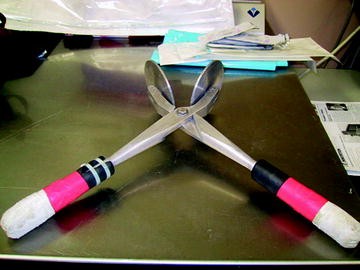
Freeze clamp.
-
2.
Place tissue(s) in the middle of the frozen clamp and quickly close the handles to flatten the sample(s), immediately followed by immersing in liquid N2 to freeze.

1.4 Protocol for TCA Extraction
-
1.
All extraction steps are carried out at 0–4°C (e.g., on ice).
-
2.
Extract frozen or lyophilized powders (e.g., 30–40 mg) in microfuge tubes with 4–6:1 or 40–60:1 (v/w) volumes of 10% (w/v) ice-cold TCA, respectively. Vortex the mixture rigorously into a homogenous suspension. If homogenization is difficult by vortexing, it can be aided by a pellet pestle for microfuge tubes (cf. Fig. 7). If frozen powder is extracted, make sure that the homogenate is completely thawed before centrifugation.
Fig. 7. 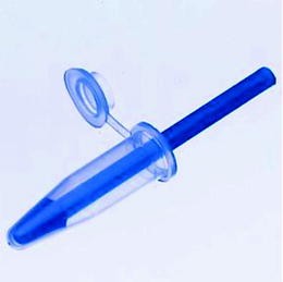
Pellet pestle for microfuge tubes.
-
3.
Centrifuge the homogenate in a refrigerated centrifuge (set at 4°C) at ≥15,000 rpm in a microfuge rotor for 20–30 min.
-
4.
Recover the supernatant with a fine-tip transfer pipette and store on ice. It is important to avoid pipetting tissue powder into the supernatant.
-
5.
Extract the residue again according to Step 2 and centrifuge according to Step 3.
-
6.
Pool supernatants from the two extractions and lyophilize using a liquid N2 pretrap to prevent TCA vapor from reaching the vacuum pump.
-
7.
Store lyophilized extract at −80°C until further analysis.

1.5 Protocol for 60% Acetonitrile Extraction
-
1.
Homogenize 5 mg of lyophilized tissue powder or cell mass in ≥0.4 mL (1:80 w/v) 60% acetonitrile (HPLC or higher solvent grade). Homogenization is achieved by rigorous vortexing or by mechanical shaking in glass beads using a micro ball mill (e.g., MM200, Retsch, Inc.).
-
2.
Incubate the homogenate for 30 min at −80°C to facilitate precipitation of macromolecules.
-
3.
Centrifuge the thawed sample at ≥15,000 g for 20–30 min at 4°C.
-
4.
Recover supernatant using a fine-tip transfer pipette into a 2-mL polypropylene microfuge tube or a 7-mL scintillation vial.
-
5.
Repeat steps 1–3 (the −80°C step can be omitted) and pool supernatants.
-
6.
Lyophilize extract using a liquid N2 pretrap to prevent acetonitrile vapor from reaching the vacuum pump.
-
7.
Store lyophilized extract at −80 C until analysis.
1.6 Protocol for Chloroform/Methanol/Water Extraction
-
1.
Homogenize 5 mg of lyophilized tissue powder or cell mass in 0.4 mL methanol in a 2-mL microfuge tube by rigorous vortexing or by mechanical shaking in glass beads for 1 min at 30 Hz using a micro ball mill (e.g., MM200, Retsch, Inc.). Alternatively, tissue or cell residue after polar metabolite extraction can be sequentially extracted with methanol for lipid components. For PCA- or TCA-extracted residues, remove acids with two to three times wash in nanopure water. All residues are lyophilized before lipid extraction.
-
2.
Incubate the homogenate for 20 min at 60 C.
-
3.
Cool and centrifuge sample at ≥15,000 g for 30 min at 4 C.
-
4.
Recover supernatant using a fine-tip transfer pipette into a screw-cap glass vial.
-
5.
Add 0.4 mL nanopure water and 0.2 mL chloroform and shake rigorously for 2 min.
-
6.
Centrifuge the biphasic solvent mixture at ≥14,000 g for 10 min at 4°C to separate the two phases.
-
7.
Recover the top aqueous layer using a fine-tip transfer pipette into a 2-mL microfuge tube or a 7-mL scintillation vial. Avoid transferring the protein pellet at the interface of the aqueous and chloroform layers.
Note: The bottom chloroform layer can also be recovered for nonpolar metabolites.
-
8.
Lyophilize extract using a liquid N2 pretrap to prevent solvent vapor from reaching the vacuum pump.
-
9.
Store lyophilized extract at −80°C until analysis.
1.7 Protocol for Methanol Extraction of Lipids
-
1.
Homogenize medium-free lyophilized cell mass or tissue powder (e.g., 1–5 mg) in 100% methanol (HPLC or MS grade) using a ratio of dry weight:methanol volume ≥100:1 plus 1 mM butylated hydroxytoluene (BHT, as antioxidant). Homogenization can be done by mechanically beating the pellet with glass beads in a micro ball mill (e.g., MM200, Retsch, Inc.) for 1 min at 30 Hz or by a pellet pestle in microfuge tubes.
-
2.
Let the homogenate stand at room temperature in the dark for 30 min.
-
3.
Centrifuge at ≥18,000 × g for 10–20 min at 4°C.
-
4.
Repeat step 1 with another aliquot of 100% methanol and centrifuge as in step 2.
-
5.
Pool both supernatants and dry under vacuum in a Speedvac or lyophilizer with a liquid N2 pretrap.
-
6.
Dissolve lipid residue in methanol or other appropriate solvents and store in Teflon-faced septum screw-cap glass vials at −80°C until analysis.
Note: Lipid extract should be filtered through 0.45-μM solvent-compatible filters (e.g., regenerated cellulose, Alltech Assoc.) prior to NMR or MS analysis.
Glossary
- BHT
-
Butylated hydroxytoluene
- CMW
-
Chloroform/methanol/water partition
- FT-ICR-MS
-
Fourier transform-ion cyclotron resonance mass spectrometry
- GC-MS
-
Gas chromatography-mass spectrometry
- GSH
-
Reduced glutathione
- GSSG
-
Oxidized glutathione
- HSQC
-
Heteronuclear single quantum coherence spectroscopy
- Isotopomer
-
Compounds of identical chemical structure but differing in isotopic composition at individual atoms
- LC-MS
-
Liquid chromatography-mass spectrometry
- MTBSTFA
-
N-methyl-N-(tert-butyldimethylsilyl)trifluoroacetamide
- PCA
-
Perchloric acid
- TCA
-
Trichloroacetic acid
- TOCSY
-
Total correlation spectroscopy
Rights and permissions
Copyright information
© 2012 Springer Science+Business Media New York
About this protocol
Cite this protocol
Fan, T.WM. (2012). Considerations of Sample Preparation for Metabolomics Investigation. In: Fan, TM., Lane, A., Higashi, R. (eds) The Handbook of Metabolomics. Methods in Pharmacology and Toxicology. Humana Press, Totowa, NJ. https://doi.org/10.1007/978-1-61779-618-0_2
Download citation
DOI: https://doi.org/10.1007/978-1-61779-618-0_2
Published:
Publisher Name: Humana Press, Totowa, NJ
Print ISBN: 978-1-61779-617-3
Online ISBN: 978-1-61779-618-0
eBook Packages: Springer Protocols