
Abstract
Diagnosis through computer-based techniques is nowadays is tremendously growing. Highly efficient system that incorporates modern techniques and fewer resources is required to speed up the diagnosis process and also to increase the level of accuracy. Fracture in a bone occurs when the external force exercised upon the bone is more than what the bone can tolerate. A disassociation between two cartilages is also referred as a fracture. The purpose of this paper is to find out the accuracy of an X-ray bone fracture detection using Canny Edge Detection method. Edge detection through Canny’s algorithm is proven to be an ideal edge identification approach in determining the end of line with impulsive threshold and less error rate.
Keywords
1 Introduction
Medical image processing is the integration of various fields that include computer science, data science, biological science and medical science. Medical image computation is an effectual and also an economic fashion that aids in the generation of visual representations of the internal part of the body for clinical inspection and medical diagnosis. To extricate the information which is clinically important from the medical image is the principal objective of medical image processing. The field of medical imaging has been witnessing advances not only in acquisition of medical images but also in its techniques and expertise of interpretation. The most common ailment of human bone is fracture. Bone fractures are nothing but the ruptures which occur due to accidents. There are many types of bone fractures such as normal, transverse, comminute, oblique, spiral, segmented, avulsed, impacted, torus and greenstick. Computer-aided diagnosis is exceedingly active field of research in which computer systems are developed to provide a quick and accurate diagnosis. A bone fracture is the outcome of intense force exertion, thrust or a slight trauma or a shock. A damaged bone is referred as a fracture. It can range from a slight crack to a completely collapsed bone, referred as hairline fracture. Generally, a bone gets fractured, if the force thrust upon it is more than what it can tolerate. The force frail the bone and breaks it. The asperity of the fracture is determined by the strength of the force. Falls, direct strikes on the body surface, traumatic events, etc., can cause bone fractures. Computerized diagnosis of fracture has helped the physician to diagnose the fracture with higher accuracy and in less time. The main objective of this work is to use the advancement of computer processing techniques to automatically diagnose the fracture. This paper focuses on the utilization of canny edge detection algorithm that helps the radiologist in automated diagnosis of image.
2 Motivation
The main reasons behind bringing up this system are:
To reduce the human faults as it is very obvious that diagnosis done by human experts can be ambiguous and can drop down beyond admissible levels. Computerized processing can be proved helpful in such case as the efficiency of software does not degrade due to overuse. Computerized system also helps to cut down the time and effort that is required to train the practitioners, and this system can be incorporated with the software of the X-ray imaging device that allows the physicians to make quick and effective diagnosis [1].
Additional inspiration behind building this system is to comfort the physicians, patients and researchers to look forward various cases for exploration.
3 Existing Methodology
Nowadays, X-ray image is widely used for fracture diagnosis and bone treatment as it provides accurate results and quick treatment. Several researches have been previously done for the same purpose. Numerous softwares had been implemented to diagnose fracture in human body. In this paper, fracture diagnosis is done using canny edge detection method. Software using canny edge detection has been implemented, and it also provide quite accurate results, but the work can be modified and more refined results can be obtained. This increases the efficiency of software. In the previous work, the edge detection operator, Sobel operator, is used for the edge identification which does not uses any sigma parameter, so the image obtained by this can be more enhanced. This work uses the Sobel operator on the value 4.75 as sigma parameter. The difference between using Sobel operator with and without parameter is shown in Fig. 1a–c.

a Original image, b edge detection using Sobel operator without sigma value, c edge detection using Sobel operator with sigma value of 4.75
4 Image Processing Technique
The technique developed here uses canny edge detection algorithm.
-
Canny Edge
Edges are considered to be most significant aspect that provides crucial information for analysis of image [2]. Edges are basically the outline that differentiates the object with its background. Identification of edge is quite a complex process that is affected by deterioration due to different fluctuating level of noise [2]. Canny edge detector is an edge detection operator. It uses multi-stage algorithm to identify broad spectrum of edges [3]. Canny Edge Detection algorithm is proven to be an optimal algorithm for edge identification. It takes a greyscale image (X-ray image) as an input, processes it and produces the output that shows the discontinuities of intensity.
The canny edge detector works in five phases: smoothing of input image by Gaussian filter, finding gradients of the image, non-maximum suppression, double thresholding and then edge tracking by hysteresis. There are several criteria on edge detecting that can be fulfilled by canny edge detection:
-
1.
Canny has a better detection (for detection criteria). Canny method has the capability to highlight all the existing edges that match with the user-determined parameter’s threshold [4].
-
2.
Canny has better localizing way. Canny is capable of producing minimum gap between detected edge and the real image edge [4].
-
3.
It provides obvious response. It gives single response for every edge. This makes less confusion on edge detection for the next image. Identifying parameters on Canny Edge Detection will give effect on every result and edge detection. The parameters are [4]:
-
(a)
Gaussian deviation standard value.
-
(b)
Threshold value.
-
(a)
The following are the steps to do Canny Edge Detection:
-
1.
Remove all noise on the image by implementing Gaussian filter. After applying Gaussian filter, the resulting image that will be obtained will be less blur. The intention of applying the filter is to obtain the real edge of the image. If the Gaussian filter is not applied, sometimes noise itself will be detected as an edge [4].
-
2.
Detect the edge with Sobel operator on the value “4.75”, and this will give the clear view of the edges and distortions. Following is an example of an edge detection using Sobel operator (Fig. 2):
Fig. 2 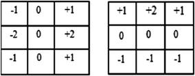
Application of Sobel operator for edge detection in horizontal and vertical directions (Gx) and (Gy)

The results from both the operators are combined to fetch the combined result by the equation given below [4]:
-
3.
Determine the direction by using the formula:
Detection uses two thresholds (maximum threshold value and minimum threshold value). If the pixel gradient is higher than maximum threshold, the pixel will be denied as background image. If the pixel gradient between maximum threshold and minimum threshold, the pixel will be accepted as an edge if it is connected with other edge pixel that is higher than the maximum threshold [4].
-
4.
Minimize the emerging edge line by applying non-maximum suppression. The process gives slimmer edge line [4].
-
5.
The last step is to fetch the binary value of the image pixels by applying two threshold values.
5 System Overview
System overview of this project is discussed through flow chart. The flow chart explains the process flow from detecting X-ray image until producing the bone fracture detection on the X-ray image (Fig. 3).

Flow chart of the system
The realization of the system is demonstrated below:
-
1.
Firstly user fed the X-ray as an input into the system. The input gets processed.
-
2.
The previous step is then carried further by filtering the input image by using filter. Here, in this system, Gaussian filter is used.
-
3.
The next step is performed after the filtered image is obtained. The image will be then processed using Canny Edge Detection method. As a result, it will produce more visible lines on the X-ray image.
-
4.
The system then couples the result of previous step with the original image. After this step, the edges of the bone get clearly visible and then further this image is processed by the system.
-
5.
To detect the location of fracture in the image, shape detection with multiple parameters is used by the system. A broken bone is expressed when the line has an end and does not have any connection with other line [4].
-
6.
The various parameters on which the system specifies the location of broken bone are as follows:
-
(a)
The red colour indicates the position where the line ends after it gets processed by canny edge detection. The line obtained is a single line.
-
(b)
The blue colour indicates the position of the line that is next to one another; such types of lines generally indicate the hairline fracture.
-
(c)
The green colour depicts the location of the end of the line with multiple ends.
-
(a)
6 Results
This system processes the input X-ray with canny edge detection method. In Fig. 4a, the user inputs the image which is an X-ray image of the bone. Then, the system will undergo canny edge detection that includes Gaussian filter and edge detection Sobel operator followed by non-maximum suppression, and the output obtained through this step is shown in Fig. 4b. In Fig. 4c, the system demonstrates the output by combining the output of the result obtained in Fig. 4b and by inverting the original image that is uploaded by the user. After this, the system detects the location where it highlights the end of line, which is shown in Fig. 4d. In Fig. 4e, the system identifies the fractured bone and location of fracture (Fig. 5).

a Input image, b output image through canny edge detection, c inverted output image by canny edge detection, d output image with canny edge detection at every edge, e output image with fracture detection

a Original image, b image after undergoing Gaussian filter, c fracture detection using Sobel operator
7 Conclusion
In this paper, fracture identification is done using Canny Edge Detection operator. This framework will help the physicians to obtain more accurate results with less effort and also in less time. The system has been tested upon real data. Using Sobel operator with the parameter sigma 4.75 helps to enhance the efficiency of the system and also helps to diagnose the hairline fracture more effectively. Because hairline fracture is basically the fracture that has multiple fractures combined together, in this fracture the complete distortion of bone occurs. So, at this value, edges can be diagnosed in such a way that all the distortions and joints are clearly visible that help to increase the success rate of the system. Along with that, soft computing techniques [5,6,7,8,9] and formal methods [10] will also be investigated to improve the clustering performance.
References
Dhanabal, R.: Digital image processing using sobel edge detection algorithm in FPGA. J. Theor. Appl. Inf. 58(1) (2013)
Mahajan, S.R., et al.: Review of an enhanced fracture detection algorithm design using X-ray image processing. IJIRSET J. 1(2) (2012)
Fazal-E-Malik: Mean and standard deviation features of color histogram using laplacian filter for content-based image retrieval. J. Theor. Appl. Inf. Technol. 34(1) (2011)
Kurniawan, S.F.: Bone fracture detection using openCV. J. Theor. Appl. Inf. Technol. 64 (2015)
Gonzalez, R.C., Woods, R.E.: Digital Image Processing, 3rd edn. (2009)
Ansari, I.A., Pant, M., Ahn, C.W.: Robust and false positive free watermarking in IWT domain using SVD and ABC. Eng. Appl. Artif. Intell. 49, 114–125 (2016)
Jauhar, S.K., Pant, M., Deep, A.: An approach to solve multi-criteria supplier selection while considering environmental aspects using differential evolution. In: International Conference on Swarm, Evolutionary, and Memetic Computing, pp. 199–208. Springer International Publishing (2013)
Ahmad, A.I., Pant, M.: SVD watermarking: particle swarm optimization of scaling factors to increase the quality of watermark. In: Proceedings of Fourth International Conference on Soft Computing for Problem Solving. NIT Silchar, Assam, India, Springer India (2015)
Zaheer, H., Pant, M., Monakhov, O., Monakhova, E.: A portfolio analysis of ten national banks through differential evolution. In: Proceedings of Fifth International Conference on Soft Computing for Problem Solving, pp. 851–862. Springer, Singapore (2016)
Jauha, S. K., Pant, M.: Recent trends in supply chain management: a soft computing approach. In: Proceedings of Seventh International Conference on Bio-Inspired Computing: Theories and Applications (BIC-TA 2012), pp. 465–478. Springer, India (2013)
Author information
Authors and Affiliations
Corresponding author
Editor information
Editors and Affiliations
Rights and permissions
Copyright information
© 2018 Springer Nature Singapore Pte Ltd.
About this paper

Cite this paper
Johari, N., Singh, N. (2018). Bone Fracture Detection Using Edge Detection Technique. In: Pant, M., Ray, K., Sharma, T., Rawat, S., Bandyopadhyay, A. (eds) Soft Computing: Theories and Applications. Advances in Intelligent Systems and Computing, vol 584. Springer, Singapore. https://doi.org/10.1007/978-981-10-5699-4_2
Download citation
DOI: https://doi.org/10.1007/978-981-10-5699-4_2
Published:
Publisher Name: Springer, Singapore
Print ISBN: 978-981-10-5698-7
Online ISBN: 978-981-10-5699-4
eBook Packages: EngineeringEngineering (R0)