
Abstract
Since the first reported use of the double-umbrella Rashkind device [1] in 1992, transcatheter paravalvular leak (PVL) closure has been performed extensively by many centers around the world.
Access this chapter
Tax calculation will be finalised at checkout
Purchases are for personal use only
References
Hourihan M, Perry SB, Mandell VS, Keane JF, Rome JJ, Bittl JA, et al. Transcatheter umbrella closure of valvular and paravalvular leaks. J Am Coll Cardiol. 1992;20:1371–7.
Kim MS, Casserly IP, Garcia JA, Klein AJ, Salcedo EE, Carroll JD. Percutaneous transcatheter closure of prosthetic mitral paravalvular leaks: are we there yet ? JACC Cardiovasc Interv. 2009;2:81–90.
Ruiz CE, Jelnin V, Kronzon I, Dudiy Y, Del ValleFernandez R, Einhorn BN, et al. Clinical outcomes in patients undergoing percutaneous closure of periprosthetic paravalvular leaks. J Am Coll Cardiol. 2011;58:2210–7.
Sorajja P, Cabalka AK, Hagler DJ, Rihal CS. Long-term follow-up of percutaneous repair of paravalvular prosthetic regurgitation. J Am Coll Cardiol. 2011;58:2218–24.
Rihal CS, Sorajja P, Booker JD, Hagler DJ, Cabalka AK. Principles of percutaneous paravalvular leak closure. JACC Cardiovasc Interv. 2012;5:121–30.
Swaans MJ, Post MC, Johan van der Ven HA, Heijmen RH, Budts W, ten Berg JM. Transapical treatment of paravalvular leaks in patients with a logistic Euroscore of more than 15%: acute and 3-month outcomes of a “Proof of Concept” study. Catheter Cardiovasc Interv. 2012;79:741–7.
Reed GW, Tuzcu EM, Kapadia SR, Krishnaswamy A. Catheter-based closure of paravalvular leak. Expert Rev Cardiovasc Ther. 2014;12(6):681–92.
Calvert PA, Northridge DB, Malik IS, Shapiro L, Ludman P, Qureshi SA, et al. Percutaneous device closure of paravalvular leak: combined experience from the United Kingdom and Ireland. Circulation. 2016;134(13):934–44.
Cruz-Gonzalez I, Rama-Merchan JC, Calvert PA, Rodríguez-Collado J, Barreiro-Pérez M, Martín-Moreiras J, et al. Percutaneous closure of paravalvular leaks: a systematic review. J Interv Cardiol. 2016;29:382–92.
Sanchez-Recalde A, Moreno R, Galeote G, Jimenez-Valero S, Calvo L, Sevillano JH, et al. Immediate and mid-term clinical course after percutaneous closure of paravalvular leakage. Rev Esp Cardiol. 2014;67(8):615–23.
Paixao A, Cilingiroglu M. Paravalvular leaks: one size (or shape) doesn’t always fit all? Catheter Cardiovasc Interv. 2016;88(4):624–5.
Goktekin O, Vatankulu MA, Tasal A, Sönmez O, Başel H, Topuz U, et al. Transcatheter trans-apical closure of paravalvular mitral and aortic leaks using a new device: first in man experience. Catheter Cardiovasc Interv. 2014;83:308–14.
Ercan E, Tengiz I, Turk U, Ozyurtlu F, Alioglu E. A new device for paravalvular leak closure. Case Report. J Geriatr Cardiol. 2015;12:187–8.
Goktekin O, Vatankulu MA, Ozhan H, Ay Y, Ergelen M, Tasal A, et al. Early experience of percutaneous paravalvular leak closure using a novel Occlutech occluder. EuroIntervention. 2016;11(10):1195–200.
Yildirim A, Goktekin O, Gorgulu S, Norgaz T, Akkaya E, Aydin U, et al. A new specific device in transcatheter prosthetic paravalvular leak closure: a prospective two-center trial. Catheter Cardiovasc Interv. 2016;88(4):618–24.
Smolka G, Pysz P, Kozłowski M, Jasiński M, Gocoł R, Roleder T, et al. Transcatheter closure of paravalvular leaks using a paravalvular leak device—a prospective Polish registry. Adv Interv Cardiol. 2016;12(2):128–34.
Davidavicius G, Rucinskas K, Drasutiene A, Samalavicius R, Bilkis V, Zakarkaite D, et al. Hybrid approach for transcatheter paravalvular leak closure of mitral prosthesis in high-risk patients through transapical access. J Thorac Cardiovasc Surg. 2014;148:1965–9.
Jelnin V, Dudiy Y, Einhorn BN, Kronzon I, Cohen HA, Ruiz CE. Clinical experience with percutaneous left ventricular transapical access for interventions in structural heart defects. A safe access and secure exit. J Am Coll Cardiol Interv. 2011;4:868–74.
Smolka G, Pysz P, Jasinski M, Gocoł R, Domaradzki W, Hudziak D, et al. Transapical closure of mitral paravalvular leaks with use of amplatzer vascular plug III. J Invasive Cardiol. 2013;25(10):497–501.
Acknowledgments
We are indebted to the staff of the catheter lab at Vilnius University, Lithuania, for their help during the procedures. We are extremely grateful to Lina Puodziukaite and Rokas Simakauskas for their efforts in data collection (both are medical students in Vilnius University). The authors also thank the engineering and marketing staff at Occlutech for illustrations, for technical descriptions of the device, and for their continuing logistic support.
Conflict of Interest Statement
Dr. Eustaquio Maria Onorato is a Consultant for Occlutech, manufacturer of the device, and Co-Principal Investigator of the international, multicenter follow-up study to monitor the efficacy and safety of the Occlutech® paravalvular leak device (PLD) in patients with mitral or aortic paravalvular leaks (see Appendix).
The remaining authors have no conflicts of interest to declare.
Cases Illustrations
-
1.
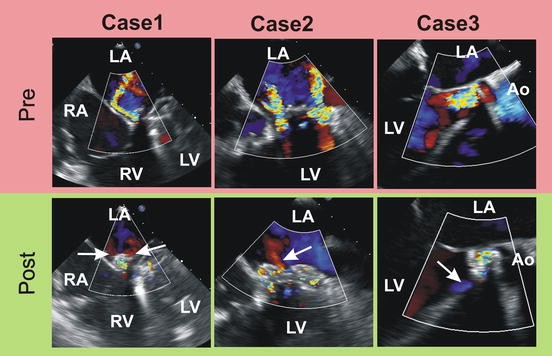
Preprocedural (red area) and postprocedural (green area) color Doppler TEE images for three cases of PVL closure. Case 1, Case 2, and Case 3 show the single mitral, double mitral, and aortic PVL closure accordingly. Postprocedural images show only trace residual regurgitant jets (arrows).
TEE transesophageal echocardiography, LA left atrium, LV left ventricle, RA right atrium, RV right ventricle, Ao aorta
-
2.
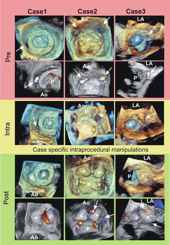
The same three cases are presented by TEE images during the procedure. Preprocedural (red area) 3D images and 3D color Doppler images cropped at level of the vena contracta clearly identified the single mitral paraprosthetic defect at 2 o’clock position in Case 1, the two mitral PVLs at 10 and 2 o’clock position in Case 2, and the aortic PVL at the noncoronary cusp projection in Case 3 (arrows). The first intraprocedural step—the wire crossing the PVL hole (arrows)—is nicely seen on 3D images in all cases (yellow area).
As the results of the following case-specific intraprocedural manipulations, all PVLs were closed (green area) with only trace residual regurgitant jets (arrows). The occluders are marked by stars, and the final orientation of the occluders is clearly seen. The occluder position is strongly parallel to the prosthesis edge in Case 1. In Case 2 the right one occluder was fixed perpendicularly because this orientation accomplished the best result. In Case 3 the occluder position is oblique.
TEE transesophageal echocardiography, P valve prosthesis, Ao aorta
-
3.
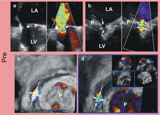
The 2D and color Doppler TEE images show the principles of the PVL measurements (arrows) in the case of flat (panel a) and tunnel (panel b) shape of the hole. The precise measurements (arrows) of the PVL performed by the cropping of regurgitant jet volume on 3D color Doppler TEE image (panel b) or using the multiplane reconstruction of same regurgitant jet volume at the level of the hole (panel b).
TEE transesophageal echocardiography, P valve prosthesis, LA left atrium, LV left ventricle
-
4.
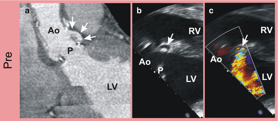
The “U”-shaped tunnel-like aortic PVL (arrows) is seen on computed tomography (panel a) and transgastric 2D TEE image (panel b). The flow through the tunnel (arrow) is detected by color Doppler in the same TEE probe position (panel c).
TEE transesophageal echocardiography, P valve prosthesis, LV left ventricle, RV right ventricle, Ao aorta
-
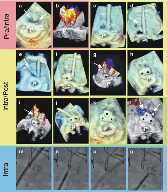
5.
The case of transapical transcatheter closure of complex mitral PVL illustrating how extremely helpful is the real-time 3D TEE during the procedure. The case depicted by pre-, intra-, and postprocedural 3D and 3D color Doppler TEE images (red, yellow, and green areas) and by cardiac fluoroscopy images (blue area). The two quite close located leaks (arrows) and two regurgitant jets were detected before the procedure (panel a and b). The first wire crossed the right leak (panel c). The second wire (arrows) fell in to the same leak and finally was placed into the left leak (panel d and e). The left leak was closed by the first occluder, a RW 8 × 4 mm (panel f and m) with a significant residual jet (arrow on the panel g). The right leak was completely closed by the second occluder, a SW 5 mm (panel h and n). A residual jet from the left leak was still observed (panel i). The third wire was passed surrounding the left leak (panel j and o), and the third occluder, a ST 5 mm, was implanted (panel k and r). Only two trace residual jets (arrows) were present after the procedure (panel l).
TEE transesophageal echocardiography, P valve prosthesis, asterisk occluder, RW rectangular waist, SW square waist, ST square twist
-
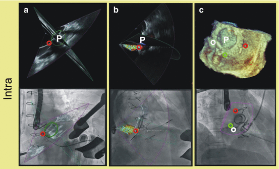
6.
The fusion of cardiac fluoroscopy and different TEE modalities.
(a)—The multiplane TEE image (on top) with red point dropped on the place of PVL is fused in the space with fluoroscopic image (on bottom). The green oval depicts the mitral prosthesis position. The wire is directed to the red point on the image.
(b)—The multiplane color Doppler image (on top) fused with fluoroscopy (on bottom) helps to visualize the PVL position signed by red point.
(c)—The fusion image of 3D TEE image shows the right position of all three PVLs. The wire is crossing the one of them signed as green point.
TEE transesophageal echocardiography, P valve prosthesis


Author information
Authors and Affiliations
Corresponding author
Editor information
Editors and Affiliations
Appendix
Appendix
The countries, institutions, and investigators participating in the international, multicenter follow-up study to monitor the efficacy and safety of the Occlutech® paravalvular leak device (PLD) in patients with mitral or aortic paravalvular leaks are listed here.
4.1.1 Coordinating Principal Investigators
Dr Eustaquio Maria Onorato, Cardiovascular Department, Humanitas Gavazzeni, Bergamo, Italy, and Prof Shakeel Ahmed Qureshi, Evelina Children’s Hospital, London, UK.
4.1.1.1 Italy
-
Humanitas Gavazzeni, Bergamo (Dr EM Onorato, Dr A Pitì, Dr F Santoro, Dr M Pennesi)
-
Centro Cardiologico Monzino, Università degli Studi di Milano (Prof. AL Bartorelli, Prof. F Alamanni, Dr G Tamborini, Dr M Muratori)
-
Clinica San Gaudenzio, Novara (Dr G Martinelli, Dr G. Cerin, Dr G Carosio, Dr M Diena)
-
Clinica San Michele, Maddaloni (Dr A De Bellis, Dr P Landino)
-
Clinica Montevergine, Mercogliano (Av) (Dr T Tesorio, Dr M Agrusta, Dr E Mango)
-
Città di Alessandria, Policlinico di Monza (Dr M Fabbrocini, Dr P Cioffi)
-
Azienda Ospedaliera SS. Antonio e Biagio e Cesare Arrigo, Alessandria (Dr M Reale, Dr M Vercellino, Dr AM Costante, Dr G Pistis, Dr D Mercogliano)
-
Dipartimento di Malattie Cardiovascolari, Campobasso (Dr CM De Filippo, Dr P Spatuzza, Dr E Caradonna)
-
USVD Emodinamica, Spedali Civili di Brescia (Dr F. Ettori, Dr S. Curello)
4.1.1.2 Lithuania
-
Santariskiu Klinikos, Vilnius (Dr A Aidietis, Dr A Zorinas, Dr Vilius Janusauskas, Dr Kestutis Rucinskas, Dr D Zakarkaite, Dr R Kramena, Dr V Bilkis)
4.1.1.3 France
-
Centre Hospitalier Universitaire, “Charles Nicole,” Rouen (Prof P-Y Litzer, Prof H Eltchaninoff, Dr M Godin, Dr F Bauer, Prof F Doguet)
-
Centre Hospitalier Universitaire, Hôpital Arnaud de Villeneuve, Montpellier (Prof B Albat, Dr T Grandet, Dr JC Macia, Dr F Cransac)
-
Centre Hospitalier Régional Universitaire, Lille (Prof F Godart, Prof F Juthier)
4.1.1.4 Cyprus
-
American Heart Institute, Nicosia (Dr C Christou, Dr S Constantinides, Dr M Neofytou)
4.1.1.5 Hungary
-
National Heart Center, Budapest (Prof A Szatmari, Dr G Fontos)
4.1.1.6 Romania
-
Centru Cardiovascular Monza, Bucharest (Prof S Balanescu, Dr A Linte)
4.1.1.7 UK
-
Castle Hill Hospital, Cottingham (Dr K Aznaouridis, Dr K Masoura, Dr D Ngaage)
Rights and permissions
Copyright information
© 2017 Springer Nature Singapore Pte Ltd.
About this chapter
Cite this chapter
Onorato, E.M. et al. (2017). Occlutech® Paravalvular Leak Device (PLD). In: Smolka, G., Wojakowski, W., Tendera, M. (eds) Transcatheter Paravalvular Leak Closure. Springer, Singapore. https://doi.org/10.1007/978-981-10-5400-6_4
Download citation
DOI: https://doi.org/10.1007/978-981-10-5400-6_4
Published:
Publisher Name: Springer, Singapore
Print ISBN: 978-981-10-5399-3
Online ISBN: 978-981-10-5400-6
eBook Packages: MedicineMedicine (R0)