
Abstract
External fixation is a method of treating bone and joint injuries as well as correcting skeletal deformities by attaching bones to an external device that stabilizes the injured limb. Additionally, it allows manipulation of the limb segments to achieve restoration of the bone’s length and alignment. Synonyms of “external fixation” are “transosseous osteosynthesis,” “external fixing,” and “external osteosynthesis” as opposed to “internal fixation”, i.e. to an osteosynthesis with the use of internal fixators (plates, nails, screws).
This is a preview of subscription content, log in via an institution.
Buying options
Tax calculation will be finalised at checkout
Purchases are for personal use only
Learn about institutional subscriptionsAuthor information
Authors and Affiliations
Corresponding author
Editor information
Editors and Affiliations
Appendixes
Appendixes
1.1 Appendix A: Dressings
Dressings are changed by a nurse. You should do this yourself only after receiving permission and proper training. The necessary materials are (Fig. 34.10):

A dressing set
-
1.
Basin or a rubber sheet
-
2.
Pair of sterile gloves
-
3.
Tweezers
-
4.
Sterile scissors
-
5.
10-ml syringe
-
6.
70% ethyl alcohol (prepared as shown in Fig. 34.11) or a chlorhexidine gluconate solution (available from a pharmacy)
Fig. 34.11 
Preparing 70% ethanol. Add 3 ml of sterile water in a syringe to 7 ml of 98% ethanol. Mix well
-
7.
3% solution of hydrogen peroxide
-
8.
5 × 5 cm sterile napkins
-
9.
Clean frame cover

Sequence of actions:
-
1.
Remove the cover from the external fixation device.
-
2.
Place the basin or spread the rubber sheet under the frame.
-
3.
Wash your hands with bactericidal soap or with antiseptic gel; put on the sterile gloves.
-
4.
Clean the frame with a sterile napkin moistened with 3% hydrogen peroxide. Then wipe the frame with a napkin moistened with chlorhexidine.
-
5.
Wipe your hands with chlorhexidine.
-
6.
Move aside the disc clips and throw away the soiled napkins (Fig. 34.12).
Fig. 34.12 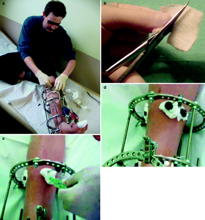
Changing the dressing. (a) Put on sterile glovers before changing the dressing. (b) Cut a 5 × 5 cm sterile napkin in half. (c) Place the napkin on the wire making use of the napkin slit. (d) Slightly press the napkin into place using the disc clips
-
7.
Using 3% hydrogen peroxide, clean the skin near the transosseous elements.
-
8.
Wipe the skin near the transosseous elements with 70% ethanol or chlorhexidine.
-
9.
Impregnate the napkins with 70% ethanol or chlorhexidine and slightly press them onto the skin using the disc clips (Fig. 34.12). Do not wrap the napkins around wires and half pins!
-
10.
Place a clean, ironed cover over the frame.

Consult your attending doctor about the frequency of dressings! Usually, if there is no suppuration and the napkins remain dry, the dressing should be changed once every 7–10 days. Simultaneously, replace the frame cover with a clean one. If the napkins get wet they must be changed—even every day—to avoid their contamination.
1.2 Appendix B: Frame Manipulation
To move the external supports of the frame, you will need two 10-mm wrenches to rotate the nuts on the connecting rods and hinges for compression, distraction, etc. (Fig. 34.13).

Wrenches
Your doctor will specify which nuts need to be adjusted in order to move the bone fragments in the right direction. To control the screws, their connecting rods should have labels, which can be made for example, with 5-mm strips of tape. Every label must have the serial number of the connecting rod and an arrow showing the direction the nuts are to be turned (Fig. 34.14). Of the two nuts, one moves a support, and the other (check-nut) maintains the rigidity of the support fixation.

Labels of the connecting rods, nuts, and check-nuts
One full turn (360o) of the nut causes a 1-mm movement of the support along the rod. Turning the nut at 90o corresponds to a support movement of 0.25 mm. The number of 0.25 mm-cycles can vary from 1 to 8 times a day, and it may be different for each of the connecting rods. To control that the nut has been turned, it could, for example, be marked.
To move a support 0.25 mm, follow this procedure: Using a wrench, move the check-nut away from the support by 0.5–1 mm. Then the nut is turned 90o. This nut is located at the site of the label. To measure a 90o turn, it is convenient to use the right angle of a corner of a sheet of paper (Fig. 34.15). The check-nut is then tightened to stabilize the support. The rest of the connecting rods should be adjusted in the same way.

Turning the nut 90°; (a) initial position, (b) nut rotated by 90°
The number of nut turns for all connecting rods will be determined by your doctor. If the recommended distraction rate is 0.25 mm four times per day, these manipulations are best performed at 8:00 AM, 12:00 PM, 04:00 PM and 08:00 PM.
Throughout the treatment period, the compression (distraction) rate may vary. It is therefore necessary to document all procedures in your orthopedic status diary, as shown in Table 34.2.
Note: It is necessary once a week (for example, on Monday) to check strength of fixing of all device nuts, except for the nuts fixing wires.
1.3 Appendix C: Your Orthopedic Status Diary
Your measurements of weight-bearing, limb circumference, and range of joint motion are critically important for your doctor. These parameters enable him or her to evaluate the functional recovery of the operated limb and to provide appropriate recommendations for your rehabilitation. Accordingly, you must enter these data on a weekly basis in the form of a special table as shown in Table 34.3.
To measure weight-bearing (Fig. 34.16), first measure your own weight by stepping onto a scale with the healthy leg. Then step on the scale using your operated leg and load it until a feeling of discomfort (pain, tension on the soft tissue, etc.) is noted. Take note of these feelings and do not overload the leg when walking.

Measuring weight-bearing: (a) measuring body weight; (b) measuring maximum possible limb loading
To measure the limb circumference, you will need a measuring tape (Fig. 34.17a). Generally, measurements are taken at three levels: at the level of the damage, and above and below it. For comparisons, the circumferences at symmetrical levels of the healthy limb are measured before treatment. Your doctor will specify the exact levels of measurement.

Measuring the circumference of the lower leg at three levels using a measuring tape (a). Measure the circumference above the damaged area of bone (b), at the level of bone damage (c), and below the area of damaged bone (d)
To control ROM, you need a protractor (Fig. 34.18). The accuracy of your measurements will be checked by your doctor.

Protractor
1.4 Appendix D: Walking with the Aid of Crutches or a Cane
1.4.1 Using Crutches
The proper use of crutches requires both good balance and coordinated movements, as well as muscle strength. Before you need to walk on crutches, learn how to use them properly and safely. This takes only a small amount of practice.
1.4.2 Walking on Crutches with Complete Unloading of the Operated Leg
Initial position: standing on the healthy leg. The crutches are positioned 6 cm forward and 15 cm laterally to your feet (Fig. 34.19).

Initial position when walking with crutches
Place the crutches 25–35 cm ahead of you. Lean on the crutches. Transferring your body, lean on your healthy leg, placed at a distance of 25–35 cm in front of the crutches. Use your hands to firmly press the upper part of the crutches to the chest without causing any pressure on the axilla. Use only your hands for support (Fig. 34.20).

Walking at partial loading with the help of crutches. White shoe print Healthy left leg, yellow shoe print operated right leg
Note: If the step of the healthy foot is shorter than the one taken by the operated leg, the next step of the operated leg should be shorter. The initial goal is to take small steps of equal length. Pay particular attention to this! Try going upstairs with the healthy leg, and downstairs with the operated one.
1.4.3 Using a Cane
Note: When placing the cane on the side of the operated leg, the load on the joints will increase by means of the body swing. The cane should be placed opposite to the damaged leg.
The first step is taken using the damaged leg. At the same time, the cane is moved ahead. When leaning on the damaged leg you should support it/yourself with the cane. Then the healthy leg is placed between the cane and the operated one. At first, the steps must be of short (shorter than the foot length) but equal length. Then move on to walking by taking steps of usual length.
You can use a cane to walk effectively if you are able to firmly hold its handle and have sufficient upper limb strength such that you do not feel pain in the joints. Using the cane outside the house will make pedestrians aware that you may be in need of assistance or require more space. A special winter cane is available for safe walking under icy street conditions. By pressing a button on the handle, a pin appears at the bottom of the cane to increase traction and prevent slippage.
Hold the cane in the hand opposite to the damaged leg (Fig. 34.21). This will result in the transfer of part of your weight to the cane, increasing the area of body support and thereby providing the body balance and confidence needed during walking. Lean on the cane at the same time that you take a step using the operated leg. This way of walking unloads the operated leg and prevents the pelvis from swinging. The use of two canes will make it easier to maintain body weight on both sides.

Cane positioning while walking
1.5 Appendix E: Clothing Adjustments
Correctly adjusted clothes will not only help you to camouflage the frame but will also provide the operated leg with warmth and ventilation and protect the insertion sites of the transosseous elements from dirt and infection.
You should wear loose-fitting clothes with a broadened sleeve or trouser leg. Pants can be split along the inner or external seam, with a triangular fabric insert placed into the slit together with a zipper or strip of Velcro (Fig. 34.22a).


Clothes, covers, and foot-hammocks. (a) Adjusted trousers; (b) the frame cover for the forearm; (c) frame cover and foot-hammock; (d) heat-insulated cover; (e) the template for the foot-hammock; (f) a slipper-based foot-hammock (g) a toe-hammock
Prepare at least two frame covers made of breathable cotton. The cover should not stretch like a stocking or trouser leg. Its length should not cover the fingers of the hand nor the foot. At the upper and lower edges of the cover insert laces so that you can fasten it above and below the frame. On the front of the cover, place a zipper, strip of Velcro, or buttons (Fig. 34.22b, c). During cold times of the year, use an additional cover made of warm material (Fig. 34.22d).
If the ExFix device is on the lower leg, use a foot-hammock to ensure that the foot position is at a right angle to the axis of the tibia, which will avoid the development of pes equinus (tip foot). To make the foot-hammock, draw a rectangular template corresponding to the size of the forefoot, as shown in Fig. 34.22e. The foot-hammock can be made from a piece of plywood 3–5 mm thick and containing drill holes in its corners to attach elastic bands (rubber cords). These should be fixed to the upper ring of the external fixation device (Fig. 34.22c).
You can wear polyurethane foam slippers, as they are light, durable, flexible, and hygienic. Some models of slippers simulate the arch of the foot. Place a rubber cord under the front part of the shoe sole (Fig. 34.22f) and fix it on the frame with tension.
Note: You should not use a hard foot-hammock in the form of the sole, as it will hinder proper gait.
Some patients have made their own crutches, even decorating them (Fig. 34.23).

(a, b) Creative crutch designs
1.6 Appendix F: Isometric Exercises
The basis of isometric exercise is muscle tension without joint motion. This is achieved either by resting the foot (hand) against an immobile support or by muscle straining, as if to exhibit well-defined abdominal muscles.
Regularly carrying out isometric exercises will improve your postoperative rehabilitation. The exercises should be done several times a day for 10 min each time. You will soon feel positive changes, including an increase in the tone and strength of the muscles.
The basic guidelines of isometric exercises are:
-
Exercise with maximum effort; strain and relax muscles smoothly.
-
Breathe rhythmically (inhale 6 s, exhale 6 s), straining the muscles during exhalation.
-
Do each exercise 6–10 times, then pause for 30–60 s before repeating the exercise.
For the first 2 months, do 9–12 exercises, then exchange 3–6 of the exercises for new ones, adding another three each month until you have a set of 20–24 exercises per training session.
Among the following exercises, your doctor will choose the best ones for you. You should determine your load (number of exercises) based on the advice of your physician or exercise therapist.
1.6.1 Isometric Exercises for Upper Limb Injuries
-
1.
Hold out your arms and place your bent fingers against a table. Breathe in; then, while breathing out, press your fingers on the table as if you want to push it through the floor.
-
2.
Bend your arms, make a fist, and press it against the table with effort, as if you are trying to push it away.
-
3.
Place your hands under a table-top and push up, trying to lift it.
-
4.
Set your arms behind the back of a chair and try to lean forward, despite the resistance of your hands.
-
5.
Hold the seat of your chair with your hands and try to lift yourself.
-
6.
Stand behind a chair, hold its back, and try to alternately compress and stretch it, as if you are playing the accordion.
-
7.
Lean your elbows on a table, pressing your hands against each other on your forehead. While trying to overcome the resistance of your hands, tilt your head forward. Relax the neck and shoulder girdle muscles and repeat the exercise 1 min later.
-
8.
Lean your elbows on a table and tilt your head back. Press your chin on your palms, trying to lower your head.
-
9.
Join your hands behind your neck, trying to push it forward. At the same time resist using the full strength of your neck muscles.
-
10.
Stretch out your arms. One hand should make a fist, the other one should clasp the fist. Push your hand against each other. Shift hands.
-
11.
Sit on a chair, join your legs together, and put your hands under your thighs close to your knees. Try to lift your shoulders up; without bending your hands, press your palms to the underside of your thighs.
-
12.
Stand with your face to a wall at a distance of about one step from it. Raise your hands high above your head and place them against the wall, trying to move it away.
-
13.
Extend your hands forward, palms inward. Press your palms against each other in full strength. Turn your palms outward and push again.
-
14.
Extend your hands forward and join the fingers together. Without releasing your fingers, try to stretch your hands apart.
1.6.2 Isometric Exercises for Lower Limb Injuries
-
1.
Lying or sitting, strain the muscles in your buttocks.
-
2.
In the supine position, strain the thigh muscles, trying to move the kneecap upwards. Repeat with the other leg.
-
3.
Sitting on a chair, clasp its legs using your feet. Straining all your leg muscles, try to press the chair.
-
4.
Sit on a chair. With raised and straightened legs, put your hands on your thighs. Press your hands on your legs and try to lift both legs without bending your knees.
-
5.
Lying on a bed, place your feet on its back. Pushing against it, stretch your feet as if you are trying to stand on tiptoes.
-
6.
Strain the sural muscle of one leg. Repeat with the other leg.
1.7 Appendix G: Rehabilatory Gymnastics
Two sets of physiotherapy exercises are presented in Tables 34.4 and 34.5 (Fig. 34.24).

(a–d) Exercise for the development of movements in the shoulder joint: creeping with your fingers
An electronic version of this brochure can be found at http://rniito.org/solomin/download/forpatient-engl.pdf and http://ortho-suv.org
Rights and permissions
Copyright information
© 2012 Springer-Verlag Italia
About this chapter
Cite this chapter
Solomin, L.N., Vorontsova, T.N., Ershov, V.V. (2012). External Fixation: a Brochure Containing Useful Information for Patients. In: Solomin, L. (eds) The Basic Principles of External Skeletal Fixation Using the Ilizarov and Other Devices. Springer, Milano. https://doi.org/10.1007/978-88-470-2619-3_34
Download citation
DOI: https://doi.org/10.1007/978-88-470-2619-3_34
Published:
Publisher Name: Springer, Milano
Print ISBN: 978-88-470-2618-6
Online ISBN: 978-88-470-2619-3
eBook Packages: Medicine