
Abstract
This chapters covers fundamental aspects of neurointerventional procedures, including preprocedural preparation, vascular access, antithrombotic management, post-procedural care, and complication management and avoidance. The Appendix discusses the neurointerventional suite.
Access this chapter
Tax calculation will be finalised at checkout
Purchases are for personal use only
References
Mold JW, Stein HF. The cascade effect in the clinical care of patients. N Engl J Med. 1986;314:512–4.
Woolf SH, Kuzel AJ, Dovey SM, Phillips Jr RL. A string of mistakes: the importance of cascade analysis in describing, counting, and preventing medical errors. Ann Fam Med. 2004;2:317–26.
Schulz K. Being wrong: adventures in the margin of error. New York: Harper Collins; 2010.
Schwartz J. Who needs hackers? New York Times 2007 Sept 12, 2007.
Mueller C, Buerkle G, Buettner HJ, et al. Prevention of contrast media-associated nephropathy: randomized comparison of 2 hydration regimens in 1620 patients undergoing coronary angioplasty. Arch Intern Med. 2002;162:329–36.
Tepel M, van der Giet M, Schwarzfeld C, Laufer U, Liermann D, Zidek W. Prevention of radiographic-contrast-agent-induced reductions in renal function by acetylcysteine. N Engl J Med. 2000;343:180–4.
Manual on Contrast Media, Version 7. Reston: American College of Radiology; 2010.
Rosovsky MA, Rusinek H, Berenstein A, Basak S, Setton A, Nelson PK. High-dose administration of nonionic contrast media: a retrospective review. Radiology. 1996;200:119–22.
Kjonniksen I, Andersen BM, Sondenaa VG, Segadal L. Preoperative hair removal–a systematic literature review. AORN J. 2002;75:928–38, 40.
Al-Mubarak N, Vitek JJ, Iyer SS, New G, Roubin GS. Carotid stenting with distal-balloon protection via the transbrachial approach. J Endovasc Ther. 2001;8:571–5.
Bendok BR, Przybylo JH, Parkinson R, Hu Y, Awad IA, Batjer HH. Neuroendovascular interventions for intracranial posterior circulation disease via the transradial approach: technical case report. Neurosurgery. 2005;56:E626; discussion E.
Kim JY, Yoon J, Jung HS, et al. Feasibility of the radial artery as a vascular access route in performing primary percutaneous coronary intervention. Yonsei Med J. 2005;46:503–10.
Yoo BS, Yoon J, Ko JY, et al. Anatomical consideration of the radial artery for transradial coronary procedures: arterial diameter, branching anomaly and vessel tortuosity. Int J Cardiol. 2005;101:421–7.
McIvor J, Rhymer JC. 245 transaxillary arteriograms in arteriopathic patients: success rate and complications. Clin Radiol. 1992;45:390–4.
Ross IB, Luzardo GD. Direct access to the carotid circulation by cut down for endovascular neuro-interventions. Surg Neurol. 2006;65:207–11; discussion 11.
Nii K, Kazekawa K, Onizuka M, et al. Direct carotid puncture for the endovascular treatment of anterior circulation aneurysms. AJNR Am J Neuroradiol. 2006;27:1502–4.
Blanc R, Mounayer C, Piotin M, Sadik JC, Spelle L, Moret J. Hemostatic closure device after carotid puncture for stent and coil placement in an intracranial aneurysm: technical note. AJNR Am J Neuroradiol. 2002;23:978–81.
Qureshi AI, Luft AR, Sharma M, Guterman LR, Hopkins LN. Prevention and treatment of thromboembolic and ischemic complications associated with endovascular procedures: part II–clinical aspects and recommendations. Neurosurgery. 2000;46:1360–75; discussion 75–6.
Derdeyn CP, Cross 3rd DT, Moran CJ, et al. Postprocedure ischemic events after treatment of intracranial aneurysms with guglielmi detachable coils. J Neurosurg. 2002;96:837–43.
Friedman JA, Nichols DA, Meyer FB, et al. Guglielmi detachable coil treatment of ruptured saccular cerebral aneurysms: retrospective review of a 10-year single-center experience. AJNR Am J Neuroradiol. 2003;24:526–33.
Ross IB, Dhillon GS. Complications of endovascular treatment of cerebral aneurysms. Surg Neurol. 2005;64:12–8.
Saw J, Bajzer C, Casserly IP, et al. Evaluating the optimal activated clotting time during carotid artery stenting. Am J Cardiol. 2006;97:1657–60.
Castellan L, Causin F, Danieli D, Perini S. Carotid stenting with filter protection. Correlation of ACT values with angiographic and histopathologic findings. J Neuroradiol. 2003;30:103–8.
Lewis BE, Ferguson JJ, Grassman ED, et al. Successful coronary interventions performed with argatroban anticoagulation in patients with heparin-induced thrombocytopenia and thrombosis syndrome. J Invasive Cardiol. 1996;8:410–7.
Matthai Jr WH. Use of argatroban during percutaneous coronary interventions in patients with heparin-induced thrombocytopenia. Semin Thromb Hemost. 1999;25 Suppl 1:57–60.
Harrigan MR, Levy EI, Bendok BR, Hopkins LN. Bivalirudin for endovascular intervention in acute ischemic stroke: case report. Neurosurgery. 2004;54:218–22; discussion 22–3.
Clayton SB, Acsell JR, Crumbley 3rd AJ, Uber WE. Cardiopulmonary bypass with bivalirudin in type II heparin-induced thrombocytopenia. Ann Thorac Surg. 2004;78:2167–9.
Keeling D, Davidson S, Watson H. The management of heparin-induced thrombocytopenia. Br J Haematol. 2006;133:259–69.
Steinhubl SR, Berger PB, Mann 3rd JT, et al. Early and sustained dual oral antiplatelet therapy following percutaneous coronary intervention: a randomized controlled trial. JAMA. 2002;288:2411–20.
Ries T, Grzyska U, Fiehler J. Antiaggregation before, during, and after coiling of unruptured aneurysms: growing evidence between scylla and charybdis. AJNR Am J Neuroradiol. 2008;29:e33.
Hwang G, Jung C, Park SQ, et al. Thromboembolic complications of elective coil embolization of unruptured aneurysms: the effect of oral antiplatelet preparation on periprocedural thromboembolic complication. Neurosurgery. 2010;67:743–8; discussion 8.
Kang HS, Han MH, Kwon BJ, et al. Is clopidogrel premedication useful to reduce thromboembolic events during coil embolization for unruptured intracranial aneurysms? Neurosurgery. 2010;67:1371–6; discussion 6.
Yamada NK, Cross 3rd DT, Pilgram TK, Moran CJ, Derdeyn CP, Dacey Jr RG. Effect of antiplatelet therapy on thromboembolic complications of elective coil embolization of cerebral aneurysms. AJNR Am J Neuroradiol. 2007;28:1778–82.
Zhang XD, Wu HT, Zhu J, He ZH, Chai WN, Sun XC. Delayed intracranial hemorrhage associated with antiplatelet therapy in stent-assisted coil embolized cerebral aneurysms. Acta Neurochir Suppl. 2011;110:133–9.
Yan BP, Clark DJ, Ajani AE. Oral antiplatelet therapy and percutaneous coronary intervention. Expert Opin Pharmacother. 2005;6:3–12.
Siller-Matula JM, Huber K, Christ G, et al. Impact of clopidogrel loading dose on clinical outcome in patients undergoing percutaneous coronary intervention: a systematic review and meta-analysis. Heart. 2011;97:98–105.
Lee DH, Arat A, Morsi H, Shaltoni H, Harris JR, Mawad ME. Dual antiplatelet therapy monitoring for neurointerventional procedures using a point-of-care platelet function test: a single-center experience. AJNR Am J Neuroradiol. 2008;29:1389–94.
Mangiacapra F, Muller O, Ntalianis A, et al. Comparison of 600 versus 300-mg clopidogrel loading dose in patients with ST-segment elevation myocardial infarction undergoing primary coronary angioplasty. Am J Cardiol. 2010;106:1208–11.
Gruberg L, Beyar R. Optimized combination of antiplatelet treatment and anticoagulation for percutaneous coronary intervention: the final word is not out yet! letter; comment. J Invasive Cardiol. 2002;14:251–3.
Schleinitz MD, Olkin I, Heidenreich PA. Cilostazol, clopidogrel or ticlopidine to prevent sub-acute stent thrombosis: a meta-analysis of randomized trials. Am Heart J. 2004;148:990–7.
Bennett CL, Weinberg PD, Rozenberg-Ben-Dror K, Yarnold PR, Kwaan HC, Green D. Thrombotic thrombocytopenic purpura associated with ticlopidine. A review of 60 cases. Ann Intern Med. 1998;128:541–4.
Yi HJ, Gupta R, Jovin TG, et al. Initial experience with the use of intravenous eptifibatide bolus during endovascular treatment of intracranial aneurysms. AJNR Am J Neuroradiol. 2006;27:1856–60.
Ries T, Siemonsen S, Grzyska U, Zeumer H, Fiehler J. Abciximab is a safe rescue therapy in thromboembolic events complicating cerebral aneurysm coil embolization: single center experience in 42 cases and review of the literature. Stroke. 2009;40:1750–7.
Song JK, Niimi Y, Fernandez PM, et al. Thrombus formation during intracranial aneurysm coil placement: treatment with intra-arterial abciximab. AJNR Am J Neuroradiol. 2004;25:1147–53.
Steinhubl SR, Talley JD, Braden GA, et al. Point-of-care measured platelet inhibition correlates with a reduced risk of an adverse cardiac event after percutaneous coronary intervention: results of the GOLD (AU-assessing ultegra) multicenter study. Circulation. 2001;103:2572–8.
Merck & Co. Inc. Integrilin prescribing information. Whitehouse Station; 2011.
Fontana P, Dupont A, Gandrille S, et al. Adenosine diphosphate-induced platelet aggregation is associated with P2Y12 gene sequence variations in healthy subjects. Circulation. 2003;108:989–95.
Farid NA, Kurihara A, Wrighton SA. Metabolism and disposition of the thienopyridine antiplatelet drugs ticlopidine, clopidogrel, and prasugrel in humans. J Clin Pharmacol. 2010;50:126–42.
Simon T, Verstuyft C, Mary-Krause M, et al. Genetic determinants of response to clopidogrel and cardiovascular events. N Engl J Med. 2009;360:363–75.
Taubert D, von Beckerath N, Grimberg G, et al. Impact of P-glycoprotein on clopidogrel absorption. Clin Pharmacol Ther. 2006;80:486–501.
Anderson CD, Biffi A, Greenberg SM, Rosand J. Personalized approaches to clopidogrel therapy: are we there yet? Stroke. 2010;41:2997–3002.
Ellis KJ, Stouffer GA, McLeod HL, Lee CR. Clopidogrel pharmacogenomics and risk of inadequate platelet inhibition: US FDA recommendations. Pharmacogenomics. 2009;10:1799–817.
Hulot JS, Bura A, Villard E, et al. Cytochrome P450 2 C19 loss-of-function polymorphism is a major determinant of clopidogrel responsiveness in healthy subjects. Blood. 2006;108:2244–7.
Mega JL, Close SL, Wiviott SD, et al. Cytochrome p-450 polymorphisms and response to clopidogrel. N Engl J Med. 2009;360:354–62.
Sofi F, Giusti B, Marcucci R, Gori AM, Abbate R, Gensini GF. Cytochrome P450 2 C19*2 polymorphism and cardiovascular recurrences in patients taking clopidogrel: a meta-analysis. Pharmacogenomics J. 2011;11:199–206.
Administration USFaD. FDA drug safety communication: reduced effectiveness of plavix (clopidogrel) in patients who are poor metabolizers of the drug. Rockville: Administration USFaD; 2010.
Lau WC, Waskell LA, Watkins PB, et al. Atorvastatin reduces the ability of clopidogrel to inhibit platelet aggregation: a new drug-drug interaction. Circulation. 2003;107:32–7.
Nguyen T, Frishman WH, Nawarskas J, Lerner RG. Variability of response to clopidogrel: possible mechanisms and clinical implications. Cardiol Rev. 2006;14:136–42.
Feher G, Koltai K, Alkonyi B, et al. Clopidogrel resistance: role of body mass and concomitant medications. Int J Cardiol. 2007;120:188–92.
Soffer D, Moussa I, Harjai KJ, et al. Impact of angina class on inhibition of platelet aggregation following clopidogrel loading in patients undergoing coronary intervention: do we need more aggressive dosing regimens in unstable angina? Catheter Cardiovasc Interv. 2003;59:21–5.
Kanaan H, Jankowitz B, Aleu A, et al. In-stent thrombosis and stenosis after neck-remodeling device-assisted coil embolization of intracranial aneurysms. Neurosurgery. 2010;67:1523–33.
Kang H-S, Kwon BJ, Kim JE, Han MH. Preinterventional clopidogrel response variability for coil embolization of intracranial aneurysms: clinical implications. AJNR Am J Neuroradiol. 2010;31:1206–10.
Seidel H, Rahman MM, Scharf RE. Monitoring of antiplatelet therapy. Current limitations, challenges, and perspectives. Hamostaseologie. 2011;31:41–51.
Bonello L, Tantry US, Marcucci R, et al. Consensus and future directions on the definition of high on-treatment platelet reactivity to adenosine diphosphate. J Am Coll Cardiol. 2010;56:919–33.
Price MJ, Berger PB, Teirstein PS, et al. Standard- vs high-dose clopidogrel based on platelet function testing after percutaneous coronary intervention: the GRAVITAS randomized trial. JAMA. 2011;305:1097–105.
Müller-Schunk S, Linn J, Peters N, et al. Monitoring of clopidogrel-related platelet inhibition: correlation of nonresponse with clinical outcome in supra-aortic stenting. AJNR Am J Neuroradiol. 2008;29:786–91.
Prabhakaran S, Wells KR, Lee VH, Flaherty CA, Lopes DK. Prevalence and risk factors for aspirin and clopidogrel resistance in cerebrovascular stenting. AJNR Am J Neuroradiol. 2008;29:281–5.
Pandya DJ, Fitzsimmons BF, Wolfe TJ, et al. Measurement of antiplatelet inhibition during neurointerventional procedures: the effect of antithrombotic duration and loading dose. J Neuroimaging. 2010;20:64–9.
Alberts MJ. Platelet function testing for aspirin resistance is reasonable to do: yes! Stroke. 2010;41:2400–1.
Eikelboom JW, Emery J, Hankey GJ. The use of platelet function assays may help to determine appropriate antiplatelet treatment options in a patient with recurrent stroke on baby aspirin: against. Stroke. 2010;41:2398–9.
Selim MH, Molina CA. Platelet function assays in stroke management: more study is needed. Stroke. 2010;41:2396–7.
Järemo P, Lindahl TL, Fransson SG, Richter A. Individual variations of platelet inhibition after loading doses of clopidogrel. J Intern Med. 2002;252:233–8.
Bhatt DL, Scheiman J, Abraham NS, et al. ACCF/ACG/AHA 2008 expert consensus document on reducing the gastrointestinal risks of antiplatelet therapy and NSAID use: a report of the American College of Cardiology foundation task force on clinical expert consensus documents. Circulation. 2008;118:1894–909.
Ogilvie BW, Yerino P, Kazmi F, et al. The proton pump inhibitor, omeprazole, but not lansoprazole or pantoprazole, is a metabolism-dependent inhibitor of CYP2C19: implications for coadministration with clopidogrel. Drug Metab Dispos. 2011;39:2020–33.
O’Donoghue ML, Braunwald E, Antman EM, et al. Pharmacodynamic effect and clinical efficacy of clopidogrel and prasugrel with or without a proton-pump inhibitor: an analysis of two randomised trials. Lancet. 2009;374:989–97.
Ho PM, Maddox TM, Wang L, et al. Risk of adverse outcomes associated with concomitant use of clopidogrel and proton pump inhibitors following acute coronary syndrome. JAMA. 2009;301:937–44.
Kwok CS, Loke YK. Meta-analysis: the effects of proton pump inhibitors on cardiovascular events and mortality in patients receiving clopidogrel. Aliment Pharmacol Ther. 2010;31:810–23.
Bhatt DL, Cryer BL, Contant CF, et al. Clopidogrel with or without omeprazole in coronary artery disease. N Engl J Med. 2010;363:1909–17.
Juurlink DN, Gomes TM, Mamdani MM, Gladstone DJ, Kapral MK. The safety of proton pump inhibitors and clopidogrel in patients after stroke. Stroke. 2011;42:128–32.
Lanas A, Garcia-Rodriguez LA, Arroyo MT, et al. Effect of antisecretory drugs and nitrates on the risk of ulcer bleeding associated with nonsteroidal anti-inflammatory drugs, antiplatelet agents, and anticoagulants. Am J Gastroenterol. 2007;102:507–15.
Bendszus M, Koltzenburg M, Bartsch AJ, et al. Heparin and air filters reduce embolic events caused by intra-arterial cerebral angiography: a prospective, randomized trial. Circulation. 2004;110:2210–5.
Kallmes DF, McGraw JK, Evans AJ, et al. Thrombogenicity of hydrophilic and nonhydrophilic microcatheters and guiding catheters. AJNR Am J Neuroradiol. 1997;18:1243–51.
Abe T, Hirohata M, Tanaka N, et al. Distal-tip shape-consistency testing of steam-shaped microcatheters suitable for cerebral aneurysm coil placement. AJNR Am J Neuroradiol. 2004;25:1058–61.
Kiyosue H, Hori Y, Matsumoto S, et al. Shapability, memory, and luminal changes in microcatheters after steam shaping: a comparison of 11 different microcatheters. AJNR Am J Neuroradiol. 2005;26:2610–6.
Spiotta AM, Hussain MS, Sivapatham T, et al. The versatile distal access catheter: the cleveland clinic experience. Neurosurgery. 2011;68:1677–86.
Binning MJ, Yashar P, Orion D, et al. Use of the outreach distal access catheter for microcatheter stabilization during intracranial arteriovenous malformation embolization. AJNR Am J Neuroradiol. 2011. Epub ahead of print.
Engelhorn T, Struffert T, Richter G, et al. Flat panel detector angiographic CT in the management of aneurysmal rupture during coil embolization. AJNR Am J Neuroradiol. 2008;29:1581–4.
Struffert T, Richter G, Engelhorn T, et al. Visualisation of intracerebral haemorrhage with flat-detector CT compared to multislice CT: results in 44 cases. Eur Radiol. 2009;19:619–25.
Doelken M, Struffert T, Richter G, et al. Flat-panel detector volumetric CT for visualization of subarachnoid hemorrhage and ventricles: preliminary results compared to conventional CT. Neuroradiology. 2008;50:517–23.
Struffert T, Eyupoglu IY, Huttner HB, et al. Clinical evaluation of flat-panel detector compared with multislice computed tomography in 65 patients with acute intracranial hemorrhage: initial results. Clinical article. J Neurosurg. 2010;113:901–7.
Sato K, Matsumoto Y, Kondo R, Tominaga T. Usefulness of C-arm cone-beam computed tomography in endovascular treatment of traumatic carotid cavernous fistulas: a technical case report. Neurosurgery. 2010;67:467–9; discussion 9–70.
Namba K, Niimi Y, Song JK, Berenstein A. Use of dyna-CT angiography in neuroendovascular decision-making. A case report. Interv Neuroradiol. 2009;15:67–72.
Söderman M, Babic D, Holmin S, Andersson T. Brain imaging with a flat detector C-arm: technique and clinical interest of XperCT. Neuroradiology. 2008;50:863–8.
Richards BF, Fleming JB, Shannon CN, Walters BC, Harrigan MR. Safety and cost effectiveness of step-down unit admission following elective neurointerventional procedures. J NeuroInterv Surg. 2011. Epub ahead of prin.
Cardella JF, Casarella WJ, DeWeese JA, et al. Optimal resources for the examination and endovascular treatment of the peripheral and visceral vascular systems: AHA intercouncil report on peripheral and visceral angiographic and interventional laboratories. J Vasc Interv Radiol. 2003;14:517S–30.
Larson TCI, Creasy JL, Price RR, Maciunas RJ. Angiography suite specifications. In: Maciunas RJ, editor. Endovascular neurological intervention. Park Ridge: American Association of Neurological Surgeons; 1995.
Badano A. AAPM/RSNA tutorial on equipment selection: PACS equipment overview: display systems. Radiographics. 2004;24:879–89.
Samei E, Seibert JA, Andriole K, et al. AAPM/RSNA tutorial on equipment selection: PACS equipment overview: general guidelines for purchasing and acceptance testing of PACS equipment. Radiographics. 2004;24:313–34.
American Society of Interventional and Therapeutic Neuroradiology. General considerations for endovascular surgical neuroradiologic procedures. AJNR Am J Neuroradiol. 2001;22:1S–3.
Bergeron P, Carrier R, Roy D, Blais N, Raymond J. Radiation doses to patients in neurointerventional procedures. AJNR Am J Neuroradiol. 1994;15:1809–12.
Hayakawa M, Moritake T, Kataoka F, et al. Direct measurement of patient’s entrance skin dose during neurointerventional procedure to avoid further radiation-induced skin injuries. Clin Neurol Neurosurg. 2010;112:530–6.
Moskowitz SI, Davros WJ, Kelly ME, Fiorella D, Rasmussen PA, Masaryk TJ. Cumulative radiation dose during hospitalization for aneurysmal subarachnoid hemorrhage. AJNR Am J Neuroradiol. 2010;31:1377–82.
National Council on Radiation Protection and Measurements. Limitation of exposure to Ionizing radiation: recommendations of the National Council on Radiation Protection and Measurements. Bethesda; 1993. Report No.: 116.
National Council on Radiation Protection and Measurements. Radiation dose management for fluoroscopy-guided interventional medical procedures. Bethesda: 2010. Report No.: 168.
Given CA, Thacker IC, Baker MD, Morris PP. Fluoroscopy fade for embolization of vein of Galen malformation. AJNR Am J Neuroradiol. 2003;24:267–70.
National Council on Radiation Protection and Measurements. Medical x-ray, electron beam, and gamma-ray protection for energies up to 50 MeV/G (equipment design, performance, and use): recommendations of the national council on radiation protection and measurements. Bethesda; 1989. Report No.: NCRP report 102.
Mooney RB, Flynn PA. A comparison of patient skin doses before and after replacement of a neurointerventional fluoroscopy unit. Clin Radiol. 2006;61:436–41.
National Council on Radiation Protection and Measurements. Implementation of the principle of as low as reasonable achievable (ALARA) for medical and dental personnel: recommendations of the National Council on Radiation Protection and Measurements. Bethesda: The Council; 1990. Report No.: NCRP report 107.
Edwards M. Development of radiation protection standards. Radiographics. 1991;11:699–712.
Boone JM, Levin DC. Radiation exposure to angiographers under different fluoroscopic imaging conditions. Radiology. 1991;180:861–5.
Wang W, Ionita CN, Keleshis C, et al. Progress in the development of a new angiography suite including the high resolution micro-angiographic fluoroscope (MAF), a control, acquisition, processing, and image display system (CAPIDS), and a new detector changer integrated into a commercial C-arm angiography unit to enable clinical use. Proc SPIE. 2010;7622:76225I.
Binning MJ, Orion D, Yashar P, et al. Use of the micro-angiographic fluoroscope for coiling of intracranial aneurysms. Neurosurgery. 2011;69:1131–8.
Schiemann M, Killmann R, Kleen M, Abolmaali N, Finney J, Vogl TJ. Vascular guide wire navigation with a magnetic guidance system: experimental results in a phantom. Radiology. 2004;232:475–81.
Chu JC, Hsi WC, Hubbard L, et al. Performance of magnetic field-guided navigation system for interventional neurosurgical and cardiac procedures. J Appl Clin Med Phys. 2005;6:143–9.
Seppenwoolde JH, Bartels LW, van der Weide R, Nijsen JF, van Het Schip AD, Bakker CJ. Fully MR-guided hepatic artery catheterization for selective drug delivery: a feasibility study in pigs. J Magn Reson Imaging. 2006;23:123–9.
Wacker FK, Hillenbrand C, Elgort DR, Zhang S, Duerk JL, Lewin JS. MR imaging-guided percutaneous angioplasty and stent placement in a swine model comparison of open- and closed-bore scanners. Acad Radiol. 2005;12:1085–8.
Wacker FK, Hillenbrand CM, Duerk JL, Lewin JS. MR-guided endovascular interventions: device visualization, tracking, navigation, clinical applications, and safety aspects. Magn Reson Imaging Clin N Am. 2005;13:431–9.
Author information
Authors and Affiliations
Appendix: The Neuroendovascular Suite
Appendix: The Neuroendovascular Suite
This appendix addresses the design and staffing of the endovascular suite, as well as monitoring and pharmacological considerations. The objectives are to provide an introduction to residents and fellows new to the angio suite, and serve as guidelines for the fortunate experienced operators planning a new suite.
1.1 Organization and Essential Equipment
The neurointerventional suite should be dedicated to the neurointerventional service. The AHA Intercouncil Report on Peripheral and Visceral Angiographic and Interventional Laboratories produced detailed recommendations for the design and equipment needed for an interventional suite.95 Although these guidelines were not designed specifically for neurointerventional suites, they are a useful resource for planning and equipment selection. Once new equipment is installed, a medical physicist should be contracted to check out the equipment to ensure that all the specifications are met and that the image quality is adequate. In addition, annual checks by the physicist are necessary to maintain image quality and minimize radiation dose.
-
1.
Size. The procedure room should be large enough to accommodate anesthesia personnel and their equipment, as well as additional personnel and equipment that may be needed for particular procedures. For example, electroencephalography and other electrophysiology monitoring devices are required during Wada testing. The size of a typical interventional suite is at least 30 × 25 ft, or 750 square feet, with a ceiling height of 10–12 ft.95,96
-
2.
Entrances. Separate entrances for patient transportation and for personnel, usually from the control room, facilitate rapid room turnover and reduce crowding.
-
3.
Standard equipment.
-
a.
High-resolution computer monitors with PACS access to allow viewing of imaging studies.
-
b.
Usually at least two monitors should display the patient’s haemodynamic data.
-
c.
Several other computer stations should be available for anesthesia, nursing and other personnel to access medical records, protocols, electronic copies of this handbook, etc.
-
d.
Sinks for waste and for cleaning
-
e.
Telephones
-
f.
Glass-fronted storage cabinets for equipment and devices.
-
a.
-
4.
Anesthesia requirements.
The room should be equipped with oxygen, suction, gas evacuation lines, and a separate telephone for anesthesia personnel.
-
5.
Lighting.
-
a.
Overhead lights should be controlled with a rheostat to allow dimming of the room lights.
-
b.
Foot pedals to control the lights and an overhead spotlight to illuminate procedures (such as during wire and catheter shaping and arterial access site closure) are helpful.
-
a.
-
6.
Tables
-
a.
Patient table. The patient’s table should be capable of four-way motion, permitting wide excursion and pivot rotation. The table should also be able to be angled up to 30° from horizontal, to facilitate myelographic procedures and Trendelenburg position in cardiovascular emergencies. Weight capacity should be at least 500 pounds with a higher peak capacity for performing chest compression in emergency situations.
-
b.
Second table. A second table, within easy reach of the operator, is used for device preparation and placement of devices and materials for use during procedures.
-
c.
Third table. A third table is needed for procedures in which some materials, such as glue or particles for embolization, must be kept separate from the other devices.
-
a.
-
7.
Power contrast injector.
-
a.
The power injector should be capable of delivering rates up to 50 mL/s.
-
b.
Ceiling-mounted or table-mounted injectors are preferred over floor models.
-
a.
-
8.
Control room.
-
a.
Contains the console for operating the angiographic equipment.
-
b.
The control room should be spacious enough to accommodate ancillary personnel, as well as medical students and visitors (at least 130 square feet).95
-
c.
The control room window should be expansive (≥4 ft by 8 ft) should be equipped with a Venetian blind to provide patient privacy during procedures such as groin prepping and Foley catheter placement.
-
d.
The control room should also contain an image processing workstation to permit viewing, storage, and analysis of images.
-
e.
One or more computers for looking up electronic medial records, scheduling programs, protocols, the latest football scores, etc.
-
a.
-
9.
Storage space – Often not considered during room planning. There should be enough space to store plenty of device stock, both within the angiography suite and in other rooms close by. Glass-fronted storage cabinets in the suite facilitate rapid device selection during procedures. A separate, out-of-room storage space should be at least 250 square feet.95
-
10.
Power requirements
-
a.
Three-phase 220 and 440 V AC power with a minimum of 100 A per phase.95
-
b.
Emergency room power (sufficient to keep the room running for at least several minutes in a power outage).
-
a.
-
11.
Temperature and humidity must be kept within narrow limits:96
-
a.
Temperature: 72 ± 10°
-
b.
Humidity: 45% ± 15%
-
a.
-
12.
Equipment room. A separate, cooled and ventilated room traditionally contained transformers, power modules, and related equipment. The recommended size for the equipment room is 100 square feet.95
-
a.
Modern systems have much more compact generators and electronics.
-
b.
An alternative to a separate room is an alcove for electronics that can be partially closed off by sliding or swinging doors. Air supply and return vents are positioned over the electronics cabinets, with a separate thermostat for the area.
-
a.
-
13.
Data management
-
a.
Data storage. The system should be able to store at least several weeks worth of imaging data to allow for rapid comparison of studies on patients that return for urgent follow-up, including patients who have suspected vasospasm, in whom subtle caliber changes are easier to detect if prior studies are available real-time.
-
b.
A picture archiving and communication system (PACS) is a computer system that manages the acquisition, transmission, storage, distribution, display, and interpretation of medical images. Multiframe studies such as angiography will require extensive storage capacity, especially if the raw DICOM data is stored. The advantage of storing the data as it comes out of the angio suite is that it could later be processed to better see a specific lesion. The data is also embedded with positioning information that would allow for repeating specific views at a later date. A cheap solution is to only store selected subtracted images of each study, saving each like a screen-save that cannot be later processed. PACS display systems are reviewed by Badano,97 and guidelines for the acquisition of and test of PACS are discussed in depth by Samei and colleagues.98
-
a.
1.2 Angiography Equipment
Isocentric biplane digital subtraction angiographic equipment is much preferable to a single plane system. Biplane technique decreases time necessary for procedures, reduces radiation exposure, minimizes contrast dose, and offers a definite technical advantage by permitting simultaneous imaging of the anteroposterior and lateral planes. Rotational 3D angiography is also useful in defining tortuous and intricate neurovascular anatomy. Ceiling-mounted equipment, such as video monitors and power-contrast injectors, are easier to manage and are less obtrusive than floor- or table-mounted devices.
1.3 Technical Specifications
-
1.
Generator
-
a.
Rating should be a minimum of 80 kW, kV(p) ≥125, 800 mA at 100 kV(p), or 1,000 mA at 80 kV(P), with a minimum switching time of 1 ms.
-
b.
Should be a high-frequency inverter generator with a power rating of 80–100 kW.95
-
c.
Should automatically compensate for voltage fluctuation.
-
d.
Should be spatially and electrically isolated, and high-voltage cables need to be shielded.
-
a.
-
2.
X-ray tube
-
a.
Focal spot indicates the size of the x-ray source. The smaller the source, the greater the resolution. Although some vendors only offer two, three focal spot sizes are preferred:
-
i.
Small, 0.3 mm, kilowatt rating 20–30 kW.
-
ii.
Medium, 0.8 mm, kilowatt rating 40–60 kW.
-
iii.
Large, 1.3 mm kilowatt rating 80–100 kW.
-
i.
-
b.
Heat capacity should be at least 800,000 to 1 million units.95
Anode heat unit capacity of 2.4 million heat units is typical at the present time; a unit with too few heat units will cause a lengthy procedure to be intermittently placed on standby.
-
a.
-
3.
The target angle should be ≤12°.
-
4.
The anode disk typically consists of graphite, with a surface coating of tungsten/rhenium alloy, and a minimum diameter of 150 mm.
-
5.
Image intensifier (II) size should range from 9 to 12 in. The smaller the II, the higher the resolution at high magnification (drawback: the smaller the II, the smaller the field of view). The field-of-view size can be electronically adjusted using tableside controls. The II should be selected for high resolution and high contrast ratio
-
a.
The conversion efficiency factor should be greater than 250 candelas per meter squared per milliroentgen per second (cd/m2)/mR/s measured at 80 kW.95
-
b.
Spatial resolution is measured at the II tube and should be at least 2.2 line-pairs/mm in the 9 in. field of view, and 3.3 line-pairs/mm for a 6 in. field of view.
-
c.
Contrast ratio should be at least 20:1.
-
a.
-
6.
Flat panel technology can replace the imaging intensifier. With the appropriate software, it allows for rotational cone-beam 3-D CT, allowing visualization of soft tissues just like a standard CT scan.
-
a.
Advantages: No need for correction for magnetic field distortion; radiation exposure can be reduced with acceptable image quality; larger and more variable field of view; improved image quality because flat panel detectors have a higher contrast resolution. Spatial resolution is primarily limited by the matrix size; maintains image quality longer in the life of the equipment, unlike II technology in which image quality inevitably degrades.
-
b.
Drawbacks: In the fluoroscopy mode, the displayed image is noisier than II images, which takes getting used to. More expensive to purchase. Flat panel detectors can fail and are more expensive to replace.
-
a.
-
7.
Monitors
-
a.
Five angiography video monitors are necessary and should be mounted on the ceiling. Two monitors are used to view digital subtraction angiograms or roadmaps; the other two are used for live fluoroscopic imaging during procedures. The fifth is a monitor for 3D reconstructions. Yet another monitor to display haemodynamic parameters should be positioned next to the angiography monitors. The monitors should measure at least 17 in. in the diagonal and have an antiglare coating. Large monitors are available and software can combine the equivalent of multiple displays on a single screen. Two kinds of monitors are available: CRTs and flat panels.
-
b.
Flat panel monitors
-
i.
Advantages: Good contrast and brightness. Lighter and less bulky than old cathode ray tubes.
-
ii.
Disadvantages: More expensive, may have viewing angle and noise texture limitations.
-
iii.
Minimum specs: 1,600 × 1,200 resolution, brightness ≥700 nit (cd/m2), 700:1 contrast ratio, 170° viewing angle.
-
i.
-
c.
The monitors should be able to display at least 1,024 × 1,024 resolution.99
-
d.
Spatial resolution should be at least 1.8 line pairs/mm in the 9 in. mode.
-
e.
Color display is needed for 3D imaging display, but grey-scale is used to display fluoroscopy and DSA images.
-
a.
-
8.
Vendors. All five angiography equipment manufacturers make equipment that features biplane DSA, flat panel technology, and 3-D imaging. Some advantages or disadvantages with each company are listed below:
-
a.
Toshiba
Biplane imaging equipment features a variable isocenter – allows for easier adjustments in c-arm positioning. Toshiba equipment also has a unique direct x-ray-to-electronic flat panel system, which theoretically lowers noise in the system.
-
b.
Siemens
Probably the most common biplane system worldwide. Nice 3-D imaging algorithm. Siemens x-ray tubes have a relatively high x-ray heat capacity, which makes for fewer interruptions during procedures due to over-heating. Siemens has “Dynavision,” which produces soft tissue imaging similar to CT scanning.
-
c.
Philips
Good all-around functionality. Philips’ rapid tube rotation, for 3-D imaging, produces a faster acquisition than other systems. The “default x-ray settings” on Philips equipment can produce a lower x-ray exposure for the patient, at the cost of lower imaging quality (however, these settings can be adjusted if needed).
-
d.
General Electric
Decent all-around functionality. In the past, GE has tended to have a less dense imaging matrix compared to other vendors and focal spots have been larger.
-
e.
Shimadzu
Also features a variable isocenter, and has the direct-conversion flat panel detector design. Shimadzu equipment has a unique motion-artifact correction feature. Shimadzu 3-D imaging is not as advanced as the other vendors, but is improving.
-
a.
1.4 Radiation Safety
1.4.1 Patient Radiation Exposure
-
1.
The overall radiation exposure with most neurointerventional procedures is variable, depending on the procedure.
-
a.
In a published series of eight neurointerventional cases, the mean effective radiation dose was 1.67 mSv (range, 0.44–3.44 mSv).100
-
b.
The estimated risk of death by radiation-induced cancer with the highest effective dose was approximately one for every 6,000 procedures.
-
c.
However, doses ten times that or higher can easily be reached in complicated interventional cases, with resultant hair loss.101
-
a.
-
2.
Another concern is that dose can be cumulative, and patients having multiple interventional procedures plus CT studies will be at a greater risk of skin injury or other adverse effects.102
-
3.
Although there is no defined maximum radiation exposure for patients, as the medical benefits are assumed to outweigh the presumed risks,103 radiation exposure to patients should be minimized.104
-
4.
Cornerstones of minimizing radiation dose to the patient:95
-
a.
Minimizing exposure time.
-
i.
Limit fluoroscopy time. Use the last image hold feature to study an image rather than standing on the fluoroscopy pedal.
-
ii.
Limit the pulse rate for fluoroscopy.
-
i.
-
b.
Dynamic acquisition (i.e. angio runs) gives more dose than simple fluoroscopy. Do only the angiographic runs that are needed. (e.g. use one 3D acquisition instead of six different oblique runs)
-
i.
Use as low a frame rate as necessary to see the findings.
-
ii.
Use the fluoro-fade (aka mask overlay) function instead of standard roadmapping. It uses lower dose.105
-
i.
-
c.
Collimate. It improves image quality and only exposes the area of interest.
Modern suites feature virtual collimation that allows positioning and collimation without using fluoroscopy.
-
d.
Use overhead and table-side shielding to protect body parts not being studied.
-
e.
Maximize distance from the x-ray source.
-
a.
-
5.
The National Council on Radiation Protection and Measurements (NCRP) has made recommendations for the design of structural shielding8 and x-ray equipment.106
-
6.
Have up-to-date imaging equipment. Vendors are constantly improving efficiency of their products and radiation protection protocols. A study showed maximum skin dose reduced from 4.1 to 1.0 Gy after installing a new neurointerventional suite.107
-
a.
Modern suites make use of robotic positioning that allows positioning based on position sense in the system rather than fluoroscopic positioning.
-
b.
Information on positioning and acquisition parameters is stored with each image, allowing later reproduction of views without fluoroscopic positioning.
-
c.
Advanced systems may allow spatial information imported from prior CT or MR studies guiding navigation with minimal additional radiation.
-
d.
Electronic zoom functions allow electronic magnification without additional X-ray dosage.
-
a.
1.4.2 Staff Radiation Exposure
-
1.
The National Council on Radiation Protection and Measurements has published guidelines for radiation exposure for medical personnel that are defined as “as low as reasonably achievable”.104,108
-
2.
Fluoroscopy is the major source of occupational radiation exposure.109
-
3.
The operating physician is at the greatest risk of receiving the maximum occupational dose.95
-
a.
Positioning of the x-ray tube under the table minimizes scatter radiation to the operator’s head and neck.110
-
b.
In biplane systems, keep the lateral tube on the side away from the operator.
-
a.
-
4.
Moveable, ceiling-mounted clear lead glass shields can be draped with sterile plastic and positioned over the patient, protecting the patient’s lower body and the operator from radiation exposure.
-
5.
Rolling floor mounted x-ray shields should be available to shield anesthesia or other personnel.
-
6.
Lead aprons
-
a.
Should provide at least 0.5 mm lead equivalent thickness.
-
b.
All full-time physicians, technologists, and nurses should wear custom-fitted aprons to ensure optimal coverage and comfort (Fig. 4.5).
Fig. 4.5 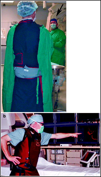
Lead apron technique. Improperly worn lead apron, as demonstrated by the junior author of this handbook early in his career (a), unfortunately witnessed by the senior author (arrow). Proper lead apron wearing technique is demonstrated by the junior author in a typically heroic pose in a more recent photograph (b).
-
c.
Extra aprons should be available for anesthesia staff and visitors.
-
a.
-
7.
Pregnant staff members.
-
a.
The NCRP-recommended maximum gestational radiation exposure is 5 mSv per gestational period, or 0.5 mSv per month.103
-
b.
Options to minimize fetal radiation exposure:
-
i.
Wear an apron with 1.0 mm lead equivalent thickness (essentially double apron coverage but they are very heavy).
-
ii.
Use draped free-standing or ceiling-mounted shields.
-
iii.
Wear wrap-around aprons to cover front and back.
-
iv.
Pregnant staff members should wear two radiation badges, with one under the apron to monitor fetal dose.
-
i.
-
a.
-
8.
Other personal radiation protection, including thyroid shields, lead glasses, should be available to all staff members.

1.5 Physiological Monitoring
Meticulous monitoring of the patient’s condition during a neurointerventional procedure is critical.
-
1.
When possible, procedures should be performed with the patient awake to permit continuous assessment of neurological status.
-
2.
Monitoring of vital signs and pulse oximetry is routine; continuous arterial line and intracranial pressure (ICP) monitoring is done as needed.
-
3.
Equipment
-
a.
Transducers should have a linear response from −10 to 400 mmHg.95
-
b.
Two or more pressure channels and two ECG channels should be available.
-
c.
An overhead monitor that projects the clinical data, including colour-coded tracings, should be positioned next to the angiography monitors.
-
d.
Arterial line monitoring. Patients with subarachnoid haemorrhage or intracranial haemorrhage should undergo continuous arterial line monitoring of their vital signs by means of a radial artery line placed before the procedure. Alternatively, monitoring may be done via the arterial sheath in patients undergoing elective procedures, such as endovascular treatment of intracranial aneurysms. To obtain an adequate tracing, the sheath must be larger than the guide catheter; monitoring can be done through a 7F femoral artery sheath when a 6F guide catheter is used.
-
e.
Intracranial pressure monitoring. Continuous monitoring of ICP should be performed in patients with a ventriculostomy. An ICP tracing on one of the overhead angiography monitors provides immediate feedback, should an abrupt change in ICP occur during the procedure, such as during an aneurysm rupture.
-
a.
1.6 Personnel
The neurointerventional team is a multidisciplinary group with expertise in neurointervention, radiology techniques, and radiation safety. At the center of this group are the interventionists, technologists, and nurses. The team is supplemented by anesthesiologists and anesthesia monitoring staff. It is important that the individuals in the team are experienced, highly motivated, and flexible. The rapidly evolving field of neurointervention, combined with the complexity of the disorders and the procedures requires that the team functions as a cohesive, adaptable unit. Moreover, the team should be large enough to allow organization of a call schedule that will provide continuous 24-h availability, without risking “burn-out” of key members.
1.6.1 Neurointerventionists
The neurointerventionist is a neurosurgeon, neuroradiologist, or neurologist who has completed a dedicated fellowship in neurointerventional radiology. Detailed knowledge of the pathophysiology of neurovascular disease must be combined with a comprehensive understanding of neuroanatomy as well as fundamentals of neurocritical care and interventional techniques.
1.6.2 Neurointerventional Technologists
Technologists in the neurointerventional suite have a background in basic radiology techniques and further expertise in computerized digital subtraction imaging. They are responsible for setting up the equipment for procedures, processing of the images, trouble-shooting during procedures, and ordering and stocking of devices. They also are responsible for alerting specialized service technicians when needed; technologists are not usually responsible for maintaining and repairing imaging equipment. At some centers, technologists may also be responsible for patient positioning and the establishment and maintenance of irrigation lines.
1.6.3 Nursing Staff
Neurointerventional nurses should be registered nurses with a background in neurointensive care. Neurointerventional nurses are responsible for patient preparation before the procedure, the establishment of intravenous access, the administration of sedation and analgesia, monitoring the patient’s condition, and maintenance of irrigation lines. Additional specific duties of the nursing staff include evaluation of the preprocedure laboratory tests, checking for allergies or drug reactions, verifying and witnessing informed consent for the procedure, placement of a Foley catheter, ventriculostomy and lumbar drain management, performance of an Allen test before radial artery procedures, monitoring peripheral pulses, and management of the arterial access site at the completion of the procedure. At some centers, a second nurse acts as the first assistant to the neurointerventionist during procedures.
1.7 Pharmacologic Considerations
Certain medications should be available for prompt access (Table 4.1). During procedures in which intravenous heparin is administered, protamine should be drawn up in a sterile syringe and placed on the back table in preparation for rapid administration in the event of a haemorrhage during the procedure. Medications used on a routine basis are listed in Table 4.2.
1.8 Future Developments
Numerous technological advances are currently in progress that offer promise in the continued evolution of the neurointerventional suite. Ultra-high resolution digital subtraction angiography has the potential to improve visualization during neurointerventional procedures. This technique has pixels sizes of 35 μm and is capable of imaging up to 30 frames per second, compared to traditional flat panel detectors, which have pixel sizes of 194 μm or larger.111,112 Magnetically guided angiography, currently available at some centers, uses extracranial magnets to help catheter and wire navigation.113,114 This technology offers the potential to permit more precise intravascular navigation while shortening fluoroscopy and procedure times. MRI-guided angiography, with the potential advantages of detailed tissue imaging as well as the elimination of radiation exposure, is another innovation under development.115–117
Rights and permissions
Copyright information
© 2013 Springer Science+Business Media New York
About this chapter
Cite this chapter
Harrigan, M.R., Deveikis, J.P. (2013). General Considerations for Neurointerventional Procedures. In: Handbook of Cerebrovascular Disease and Neurointerventional Technique. Contemporary Medical Imaging, vol 1. Humana Press, Totowa, NJ. https://doi.org/10.1007/978-1-61779-946-4_4
Download citation
DOI: https://doi.org/10.1007/978-1-61779-946-4_4
Published:
Publisher Name: Humana Press, Totowa, NJ
Print ISBN: 978-1-61779-945-7
Online ISBN: 978-1-61779-946-4
eBook Packages: MedicineMedicine (R0)