
Abstract
Objective
To develop and evaluate the performance of deep convolutional neural networks (DCNN) to detect and identify specific total shoulder arthroplasty (TSA) models.
Materials and methods
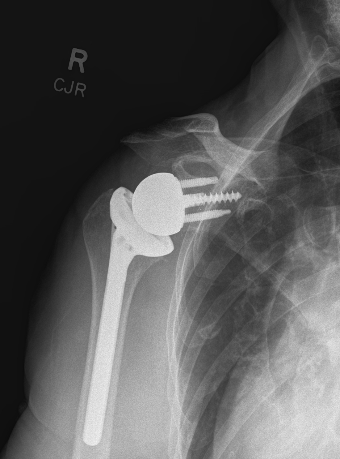
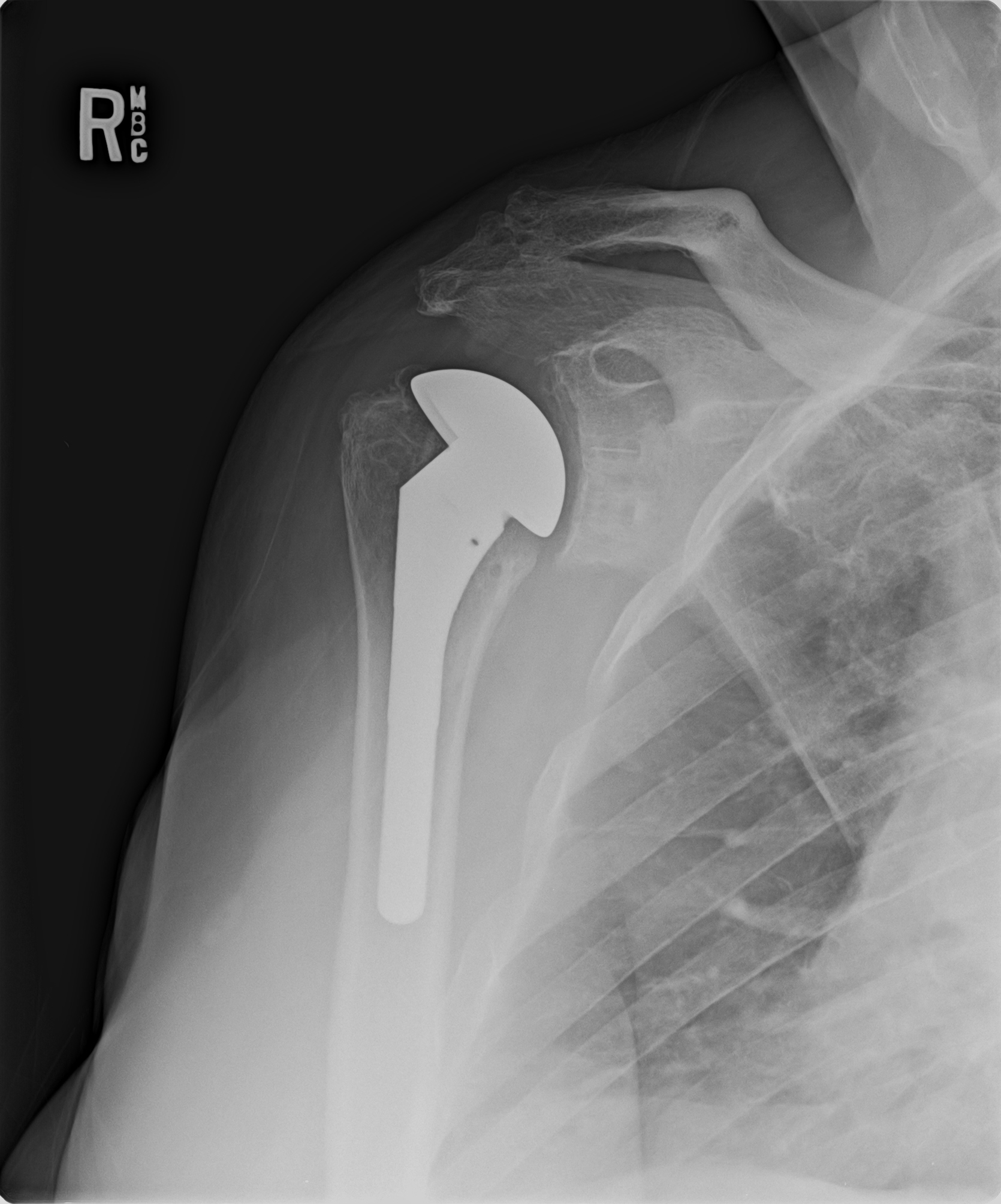
We included 482 radiography studies obtained from publicly available image repositories with native shoulders, reverse TSA (RTSA) implants, and five different TSA models. We trained separate ResNet DCNN–based binary classifiers to (1) detect the presence of shoulder arthroplasty implants, (2) differentiate between TSA and RTSA, and (3) differentiate between the five TSA models, using five individual classifiers for each model, respectively. Datasets were divided into training, validation, and test datasets. Training and validation datasets were 20-fold augmented. Test performances were assessed with area under the receiver-operating characteristic curves (AUC-ROC) analyses. Class activation mapping was used to identify distinguishing imaging features used for DCNN classification decisions.
Results
The DCNN for the detection of the presence of shoulder arthroplasty implants achieved an AUC-ROC of 1.0, whereas the AUC-ROC for differentiation between TSA and RTSA was 0.97. Class activation map analysis demonstrated the emphasis on the characteristic arthroplasty components in decision-making. DCNNs trained to distinguish between the five TSA models achieved AUC-ROCs ranging from 0.86 for Stryker Solar to 1.0 for Zimmer Bigliani-Flatow with class activation map analysis demonstrating an emphasis on unique implant design features.
Conclusion
DCNNs can accurately identify the presence of and distinguish between TSA & RTSA, and classify five specific TSA models with high accuracy. The proof of concept of these DCNNs may set the foundation for an automated arthroplasty atlas for rapid and comprehensive model identification.


References
Roberts CC, Ekelund AL, Renfree KJ, Liu PT, Chew FS. Radiologic assessment of reverse shoulder arthroplasty. RadioGraphics. 2007;27:223–35. https://doi.org/10.1148/rg.271065076.
McFarland EG, Sanguanjit P, Tasaki A, Keyurapan E, Fishman EK, Fayad LM. The reverse shoulder prosthesis: a review of imaging features and complications. Skelet Radiol. 2006;35:488–96. https://doi.org/10.1007/s00256-006-0109-1.
Dekker TJ, Steele JR, Vinson EV, Garrigues GE. Current peri-operative imaging concepts surrounding shoulder arthroplasty. Skelet Radiol. 2019;48:1485–97. https://doi.org/10.1007/s00256-019-03183-3.
Lee DH, Choi YS, Potter HG, Endo Y, Sivakumaran T, Lim TK, et al. Reverse total shoulder arthroplasty: an imaging overview. Skelet Radiol. 2020;49:19–30. https://doi.org/10.1007/s00256-019-03275-0.
Lin DJ, Wong TT, Kazam JK. Shoulder arthroplasty, from indications to complications: what the radiologist needs to know. RadioGraphics. 2016;36:192–208. https://doi.org/10.1148/rg.2016150055.
Branovacki G. Ortho atlas - hip arthroplasty - U.S. femoral implants 1938–2008: Ortho Atlas Publishing, Inc.; 2008.
IMPLANT ATLAS — Hip & Knee Book https://hipandkneebook.com/hip-implants. Accessed 15 Dec 2019.
Wilson NA, Jehn M, York S, Davis CM. Revision total hip and knee arthroplasty implant identification: implications for use of Unique Device Identification 2012 AAHKS member survey results. J Arthroplast. 2014;29:251–5. https://doi.org/10.1016/j.arth.2013.06.027.
Chea P, Mandell JC. Current applications and future directions of deep learning in musculoskeletal radiology. Skelet Radiol. 2019:1–15. https://doi.org/10.1007/s00256-019-03284-z.
Yi PH, Kim TK, Wei J, Shin J, Hui FK, Sair HI, et al. Automated semantic labeling of pediatric musculoskeletal radiographs using deep learning. Pediatr Radiol. 2019;49:1066–70. https://doi.org/10.1007/s00247-019-04408-2.
Hemke R, Buckless CG, Tsao A, Wang B, Torriani M. Deep learning for automated segmentation of pelvic muscles, fat, and bone from CT studies for body composition assessment. Skelet Radiol. 2019:1–9. https://doi.org/10.1007/s00256-019-03289-8.
Lee S, Choe EK, Kang HY, Yoon JW, Kim HS. The exploration of feature extraction and machine learning for predicting bone density from simple spine X-ray images in a Korean population. Skelet Radiol. 2019:1–6. https://doi.org/10.1007/s00256-019-03342-6.
Olczak J, Fahlberg N, Maki A, Razavian AS, Jilert A, Stark A, et al. Artificial intelligence for analyzing orthopedic trauma radiographs. Acta Orthop. 2017;88:581–6. https://doi.org/10.1080/17453674.2017.1344459.
Kim DH, MacKinnon T. Artificial intelligence in fracture detection: transfer learning from deep convolutional neural networks. Clin Radiol. 2018;73:439–45. https://doi.org/10.1016/j.crad.2017.11.015.
Lindsey R, Daluiski A, Chopra S, Lachapelle A, Mozer M, Sicular S, et al. Deep neural network improves fracture detection by clinicians. Proc Natl Acad Sci. 2018;115:11591–6. https://doi.org/10.1073/pnas.1806905115.
Radiopaedia.org, the wiki-based collaborative Radiology resource https://radiopaedia.org/. Accessed 15 Dec 2019.
Google https://www.google.com/. Accessed 15 Dec 2019.
Open-i. https://openi.nlm.nih.gov/. Accessed 15 Dec 2019.
Common US Shoulder Prostheses faculty.washington.edu/alexbert/Shoulder/CommonUSShoulderProstheses.htm. Accessed 15 Dec 2019.
Esteva A, Kuprel B, Novoa RA, Ko J, Swetter SM, Blau HM, et al. Dermatologist-level classification of skin cancer with deep neural networks. Nature. 2017;542:115–8. https://doi.org/10.1038/nature21056.
Burlina PM, Joshi NJ, Ng E, Billings SD, Rebman AW, Aucott JN. Automated detection of erythema migrans and other confounding skin lesions via deep learning. Comput Biol Med. 2019;105:151–6. https://doi.org/10.1016/j.compbiomed.2018.12.007.
Lakhani P, Gray DL, Pett CR, Nagy P, Shih G. Hello world deep learning in medical imaging. J Digit Imaging. 2018;31:283–9. https://doi.org/10.1007/s10278-018-0079-6.
Yi PH, Wei J, Kim TK, Sair HI, Hui FK, Hager GD, et al. Automated detection & classification of knee arthroplasty using deep learning. Knee. 2019. https://doi.org/10.1016/j.knee.2019.11.020.
Borjali A, Chen AF, Muratoglu OK, Morid MA, Varadarajan KM. Detecting total hip replacement prosthesis design on plain radiographs using deep convolutional neural network. J Orthop Res. 2020. https://doi.org/10.1002/jor.24617.
Russakovsky O, Deng J, Su H, Krause J, Satheesh S, Ma S, et al. ImageNet large scale visual recognition challenge 2014.
Chartrand G, Cheng PM, Vorontsov E, Drozdzal M, Turcotte S, Pal CJ, et al. Deep learning: a primer for radiologists. RadioGraphics. 2017;37:2113–31. https://doi.org/10.1148/rg.2017170077.
Soffer S, Ben-Cohen A, Shimon O, Amitai MM, Greenspan H, Klang E. Convolutional neural networks for radiologic images: a radiologist’s guide. Radiology. 2019;290:590–606. https://doi.org/10.1148/radiol.2018180547.
Cheng PM, Malhi HS. Transfer learning with convolutional neural networks for classification of abdominal ultrasound images. J Digit Imaging. 2017;30:234–43. https://doi.org/10.1007/s10278-016-9929-2.
Chung SW, Han SS, Lee JW, Oh K-S, Kim NR, Yoon JP, et al. Automated detection and classification of the proximal humerus fracture by using deep learning algorithm. Acta Orthop. 2018;89:468–73. https://doi.org/10.1080/17453674.2018.1453714.
Urakawa T, Tanaka Y, Goto S, Matsuzawa H, Watanabe K, Endo N. Detecting intertrochanteric hip fractures with orthopedist-level accuracy using a deep convolutional neural network. Skelet Radiol. 2018. https://doi.org/10.1007/s00256-018-3016-3.
Lakhani P. Deep convolutional neural networks for endotracheal tube position and X-ray image classification: challenges and opportunities. J Digit Imaging. 2017;30:460–8. https://doi.org/10.1007/s10278-017-9980-7.
Goksuluk D, Korkmaz S, Zararsiz G, Karaagaoglu A,Ergu. easyROC: an interactive web-tool for ROC curve analysis using R language environment. R J 2016;8:213. https://doi.org/10.32614/RJ-2016-042.
Incorrectly Labeled RTSA 1. https://ryortho.com/wp-content/uploads/2015/03/Zimmer_ReverseTotalShoulderReplacementXRay_WEB.jpg. Accessed 19 Apr 2020.
Incorrectly Labeled RTSA 2. http://www.jacksonorthopaedicsurgery.com/wp-content/uploads/2015/01/Monoblock_Post.jpg. Accessed 19 Apr 2020.
Incorrectly Labeled TSA 1. http://www.jacksonorthopaedicsurgery.com/wp-content/uploads/2015/01/Turon_postop.jpg. Accessed 19 Apr 2020.
Ghazala CG, Candal-Couto J. Anatomic shoulder arthroplasty. Orthop Traumatol. 2018;32:171–7. https://doi.org/10.1016/j.mporth.2018.03.005.
Lucas RM, Hsu JE, Gee AO, Neradilek MB, Matsen FA. Impaction autografting: bone-preserving, secure fixation of a standard humeral component. J Shoulder Elb Surg. 2016;25:1787–94. https://doi.org/10.1016/J.JSE.2016.03.008.
Killian ML, Cavinatto L, Galatz LM, Thomopoulos S. Recent advances in shoulder research. Arthritis Res Ther. 2012;14:214. https://doi.org/10.1186/AR3846.
False Positive 1 for Stryker Solar Classifier (Actual: DePuy Global). http://faculty.washington.edu/alexbert/Shoulder/Prostheses/Xrays/171b.jpg. Accessed 19 Apr 2020.
False Positive 2 for Stryker Solar Classifier (Actual: Biomet Biomodular) http://faculty.washington.edu/alexbert/Shoulder/Prostheses/Xrays/36i.jpg. Accessed 19 Apr 2020.
False Positive 5 for DePuy HRP Classifier (Actual: Zimmer Bigliani Flatow). http://faculty.washington.edu/alexbert/Shoulder/Prostheses/Xrays/120f.jpg. Accessed 19 Apr 2020.
False Positive 4 for DePuy HRP Classifier (Actual: Stryker Solar). http://faculty.washington.edu/alexbert/Shoulder/Prostheses/Xrays/290t.jpg. Accessed 19 Apr 2020.
False Positive 1 for DePuy HRP Classifier (Actual: DePuy Global). http://faculty.washington.edu/alexbert/Shoulder/Prostheses/Xrays/34.jpg. Accessed 19 Apr 2020.
False Positive 2 for DePuy HRP Classifier (Actual: DePuy Global). http://faculty.washington.edu/alexbert/Shoulder/Prostheses/Xrays/165c.jpg. Accessed 19 Apr 2020.
False Positive 3 for DePuy HRP Classifier (Actual: DePuy Global). http://faculty.washington.edu/alexbert/Shoulder/Prostheses/Xrays/184s.jpg. Accessed 19 Apr 2020.
False Positive for DePuy Global Classifier and False Negative for DePuy HRP Classifier (Actual: DePuy HRP). http://faculty.washington.edu/alexbert/Shoulder/Prostheses/Xrays/50.jpg. Accessed 19 Apr 2020.
False Negative 1 for DePuy Global Classifier. http://faculty.washington.edu/alexbert/Shoulder/Prostheses/Xrays/174c.jpg. Accessed 19 Apr 2020.
False Negative for Biomet Classifier. http://faculty.washington.edu/alexbert/Shoulder/Prostheses/Xrays/3c.jpg. Accessed 19 Apr 2020.
False Negative 2 for Stryker Solar Classifier. http://faculty.washington.edu/alexbert/Shoulder/Prostheses/82610579a.jpg. Accessed 19 Apr 2020.
False Negative 1 for Stryker Solar Classifier. http://faculty.washington.edu/alexbert/Shoulder/Prostheses/82610579c.jpg. Accessed 19 Apr 2020.
False Negative 2 for DePuy Global Classifier. http://faculty.washington.edu/alexbert/Shoulder/Prostheses/Xrays/39c.jpg. Accessed 19 Apr 2020.
Huang KT, Silva MA, See AP, Wu KC, Gallerani T, Zaidi HA, et al. A computer vision approach to identifying the manufacturer and model of anterior cervical spinal hardware. J Neurosurg Spine. 2019:1–7. https://doi.org/10.3171/2019.6.SPINE19463.
Lakhani P, Sundaram B. Deep learning at chest radiography: automated classification of pulmonary tuberculosis by using convolutional neural networks. Radiology. 2017;284:574–82. https://doi.org/10.1148/radiol.2017162326.
Author information
Authors and Affiliations
Corresponding author
Ethics declarations
Conflict of interest
The authors declare that they have no conflict of interest.
Additional information
Publisher’s note
Springer Nature remains neutral with regard to jurisdictional claims in published maps and institutional affiliations.
Rights and permissions
About this article

Cite this article
Yi, P.H., Kim, T.K., Wei, J. et al. Automated detection and classification of shoulder arthroplasty models using deep learning. Skeletal Radiol 49, 1623–1632 (2020). https://doi.org/10.1007/s00256-020-03463-3
Received:
Revised:
Accepted:
Published:
Issue Date:
DOI: https://doi.org/10.1007/s00256-020-03463-3